The Corticospinal Tract |
---|
The main pathway by which voluntary movements are executed is the corticospinal tract (CST). |
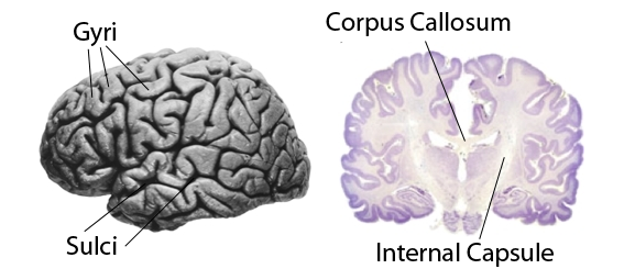
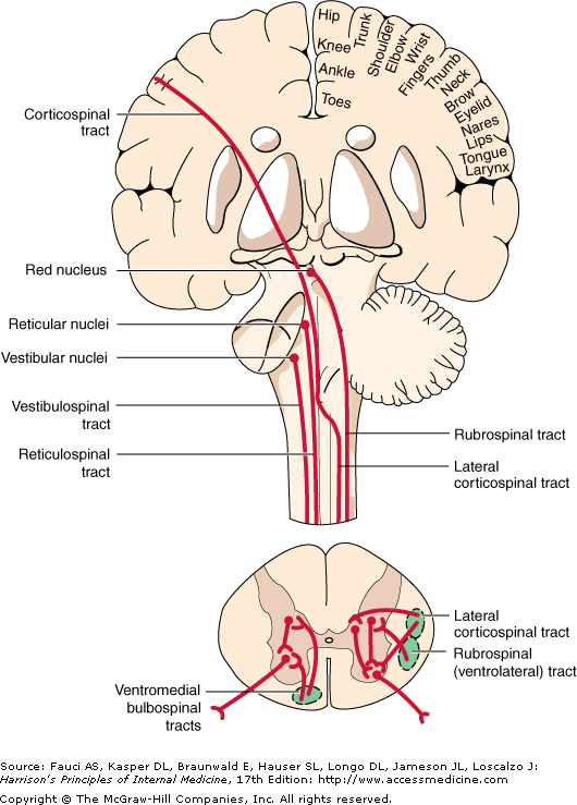
The CST begins in the pre-central gyrus of the cerebral cortex; these pyramidal cells have axons that pass along the length of the CNS to the motoneurones in the ventral horn of the spinal cord. |
The CST crosses the midline in the pyramids of the medulla: as a consequence, the left side of the brain controls movements of the right side of the body. |
A small minority (~10%) of CST axons pass down the medial part of the anterior columns of the spinal cord before crossing the midline. They control proximal muscles in the limbs. |
Within the brain, the CST passes through the Internal Capsule - between the basal ganglia and the thalamus. |
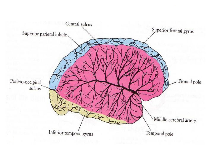
The Internal Capsule is supplied with blood by the middle cerebral artery; when this artery becomes damaged, the contralateral side of the body becomes weak or paralysed (as in a stroke). |
The Motor Cortex |
---|
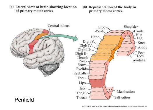
The motor cortex (the pre-central gyrus) contains an orderly map of all the possible movements of the opposite side of the body arranged in order; electrical or magnetic stimulation of each small area of motor cortex causes contractions of groups of muscles responsible for a specific movement. |
Stimulation of each area within the somatotopic map induces contractions of relevant muscle groups rather than of individual muscles. |
The corticospinal tract is the executive pathway in the control of voluntary movements, and connects the motor cortex with alpha motoneurones. |
Note that movements of the head and face are controlled by the lateral section of the pre-central gyrus. |
The central section of the precentral gyrus controls the upper limb, and a disproportionately large area of motor cortex is involved in controlling movements of the hand and fingers. |
The medial part of the precentral gyrus (including the section on the medial side of the hemisphere) controls the trunk and lower limbs |
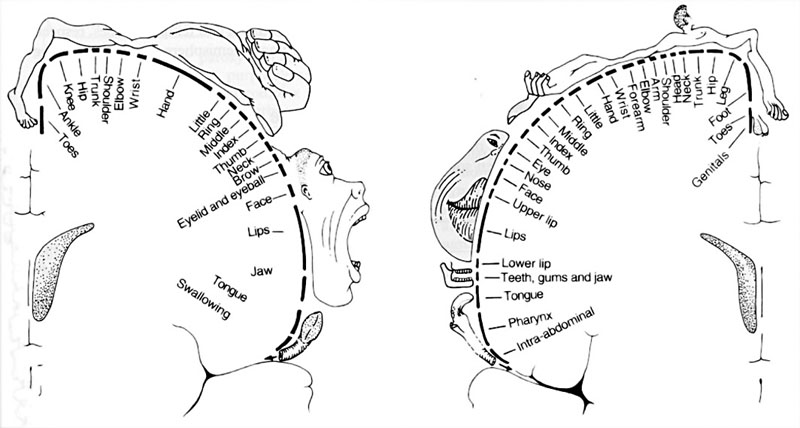
The motor homunculus is an image of the areas of motor cortex given over to movements of each part of the body |
The prehensile thumb of primates has an especially large repertoire of movements, and a very large area of motor cortex is devoted to the control of hand movements. |
The muscles of the face also have a disproportionate representation on the cortex; this is partly associated with the muscles of facial expression, but also with the muscles of the jaw and others involved in swallowing. |
The motor cortex and cortico-spinal tract are particularly concerned with producing fine, precise movements. Lesions of the motor cortex (such as the surgical ablation of one area) lead to paralysis or a loss of power in the muscles affected. In time, these muscles can regain some movement, but the movements are always coarse in comparison. |
The corticospinal tract is also called the pyramidal tract because it crosses the midline in the pyramids of the medulla. The term 'extrapyramidal' refers to pathways that can cause coarser movements, mediated by other pathways. |
The pyramidal tract, originating in the motor cortex, is sometimes referred to as the 'upper motoneurone' by clinicians. This is in contrast to the 'lower motoneurone' - i.e. the alpha motoneurones - as it is useful to distinguish between these two groups of neurones when muscles become paralysed. |
How do we know this? |
---|
Electrical stimulation of the motor cortex of anaesthetised animals provided the basic knowledge. A Canadian neurosurgeon, Wilbur Penfield, applied electrical stimuli to the human cortex and elicited movements ot eh contralateral side of the body in the sequence shown in the topographic map. |
Injuries to an area of the motor cortex cause a paralysis or weakness of the corresponding muscle groups: fine movements are particularly affected |
Modern techniques using magnetic stimuli allow stimulation of areas of motor cortex to be acheived in the conscious subject. |
Bulbospinal pathways: the Rubro-spinal, Reticulo-spinal and Vestibulo-spinal Tracts |
---|
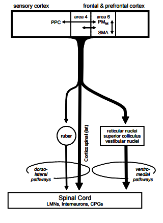
Bulbospinal pathways start in the brainstem and their axons descend through the spinal cord to reach the motoneurones. They are responsible for coarse movements and postural adjustments. |
Patients who have injuries to the motor cortex can often produce coarse movements, and cortical connections with the bulbospinal tracts (using cortico-bulbar axons) are thought to be involved in execution of these movements. |
In the cord the Rubrospinal tract, originating in the red nucleus, accompanies the corticospinal tract in the lateral columns. |
The Reticulospinal and Vestibulospinal tracts, pass down the medial part of the anterior columns of the spinal cord, and are particularly involved in muscle tone and postural responses as well as balance. |
How are the movements executed by M1 initiated and organised within the brain?
Pre-Motor Cortex and Supplementary Motor Area (Brodmann's Area 6) |
---|
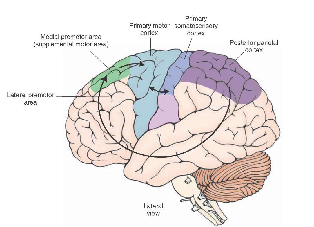
The premotor cortex (PMC, PMlat in the diagram) organises the combinations of movements that are executed by the Motor Cortex M1 (Brodmann's Area 4) in order to achieved a desired objective. |
The Supplementary Motor Cortex (SMA; M2) is medial to and has a similar role to the premotor cortex; both have similar cytoarchitecture, and comprise Brodmann's area 6. |
Electrical stimulation of PMC and SMA lead to more complex sequences of movements than stimulation of M1 |
PMC and SMA project to M1 in order to execute movements, but the some also depend on pathways from the cortex that synapse within the brainstem. These cortico-bulo-spinal paths are involved in coarse movements, unlike M1 which is concerned with fine precise movements, such as the movements of the hand and fingers. |
PMC and SMA receive inputs from the primary somatosensory cortex S1 and the posterior parietal cortex (PPC) which integrates somatosensory and visual information involved in movement. |
PMC and SMA receive inputs from the basal ganglia, which initiate movements. The basal ganglia become active ahead of any activity in PMC, SMA or M1 during voluntary movements. |
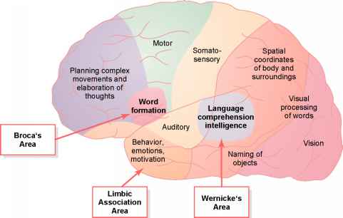
There are many motivating factors in deciding to make a voluntary movement - so many parts of the cortex are involved in different ways and in different contexts. What we know is that these areas of cortex communicate with the pre-motor and supplementary motor areas and with the basal ganglia, which is the area of the brain that initiates movement. Finally, the motor cortex and the corticospinal tract is responsible for carrying the commands to the alpha motoneurones and therefore the musculature. |
The Primary Somatosensory area (S1)lies behind the central sulcus in the post-central gyrus: it reports about proprioceptive and cutaneous events, such as the position of a limb, and the contact of the fingers with objects. |
The Posterior Parietal Cortex (PPC) lies behind S1 and integrates visual and somatosensory information concerned with spatial awareness (such as would be used in hand-eye coordination). |
PPC sends information concerning the position of objects in the visual field and of the limbs to the Pre-motor (lateral) and Supplementary Motor Cortex (medial), as well as other sites in the frontal lobe concerned with attention and thinking. These pathways may be direct using subcortical association fibres, or through the basal ganglia and thalamus.
The basal ganglia are the site in the brain where movements are initiated. They utilise information from all association areas of the cortex, such as areas of the frontal lobe involved in attention and thinking, and tactile, visual and auditory cues as to the position of the body. |
The Pre-motor and Supplementary motor areas use information from all parts of the cortex and from the basal ganglia in designing the appropriate combinations and sequences of movements that need to be executed in order to carry out any desired movement. |
The Primary Motor Cortex (M1, the origin of the corticospinal tract) receives inputs from PMC, SMA and S1 and executes the combinations and sequence of movements required to achieve the goal. |
A further input to the motor cortex comes from the cerebellum, which provides feedback - an error signal that allows the motor cortex to adjust movements and provide greater precision. |
© HumanPhysiology.Academy 2014-2015