The Human Brain

Cerebellum and Basal Ganglia

The cerebellum changed considerably as motor functions changed during the course of evolution. The earliest parts of the structure were concerned with inputs from the spinal cord - the lateral line of fish, and afferent inputs from muscles tendons and joints. The development of the vestibular organs provided a sense of balance, and one lobe of the cerebellum is devoted to the vestibular input. But the greatest expansion of the organ came with the development of the upright posture.

Structure of the Cerebellum

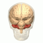
The cerebellum (red) is a solid structure attached to the back of the brainstem by two cerebellar peduncles - one on each side of the midline. These contain the axons by which the cerebellum receives information, and passes information back to the brainstem and cerebral hemispheres.

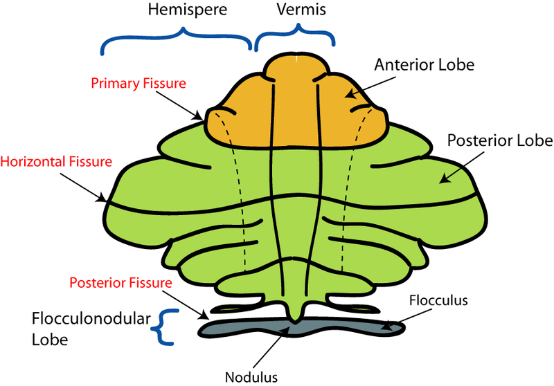
The cerebellum is divided into three lobes:

  • The Flocculonodular lobe - the oldest in evolutionary terms, and the most caudal, sometimes called the vestibulo-cerebellum, because of its association with the vestibular system
  • The Anterior lobe, sometimes called the spino-cerebellum, because of its association with spinal inputs related to proprioception
  • The Posterior lobe - the most recent development, concerned with inputs from the cortex, particularly with visual, auditory and somatosensory inputs related with body position and movement.

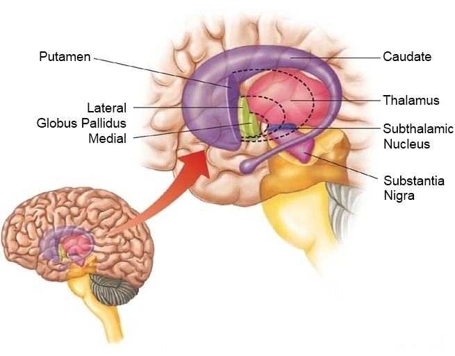
In the midline is the vermis (the nodule in the flocculonodular lobe). and laterally are the hemispheres; the area on each side of the vermis is sometimes called the intermediate lobe

Evolution of the Cerebellum

The oldest part of the cerebellum is the flocculonodular lobe, which has connections with the vestibular apparatus and is particularly concerned with balance and eye movements.

The anterior lobe has connections with the spinal cord and is concerned with coordination of the musculature.

The posterior lobe developed greatly when primates evolved and adopted the upright posture; at the same time the forebrain expanded and there are important connections between these two.

The cerebellum is concerned with the control of movement, integrating signals from different parts of the nervous system and generating error signals that allow adjustments to be made to achieve the desired objective of the movement.

In cerebellar disease, voluntary movements can become grossly exaggereated and unsuccessful attempts to correct them result in a tremor that occurs during voluntary movement.


Divisions and Nuclei of the Cerebellum
1.


The large lateral hemispheres of the posterior lobe project to the dentate nuclei, and onwards to the cerebral cortex, via the red nucleus of the midbrain

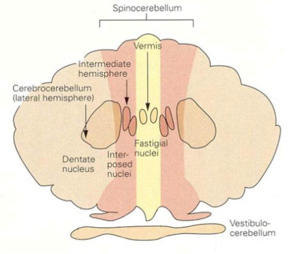
Flocculo-nodular lobe (vesibulocerebellum) projects to vestibular areas of the brainstem via the fastigial nuclei.

The interposed nuclei (N interpositus- emboliform and globose nuclei) are the output pathway for the intermediate area of cerebellar cortex, lateral to the vermis.

Connections between the Cortex and Cerebellum

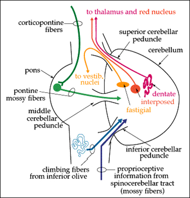
The cerebellum receives copies of the messages (motor commands) sent down the corticospinal tract.

The cerebellum compares these commands, with feedback information (information from proprioceptors, vision, touch, hearing) and decides if the command is being accomplished, and whether there are errors.

Information about any errors is returned to the cortex, so that it can adjust the command signal.

The first pathway - a copy of the command message issued by the motor cortex - involves cortico-pontine and ponto-cerebellar neurones.

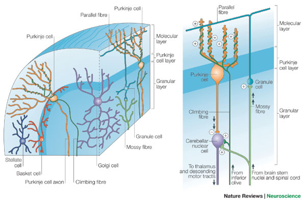
The sifting of feedback signals to extract information about errors is done by Purkinje cells, the principal cell of each cerebellar module.

The pontine fibres that feed into the cerebellum are known as 'mossy fibres' because of the ultrastructure of their terminals.

Mossy fibres supply the cerebellar cortex with information from many areas of the cortex, concerned with the position of the body, information from the visual and auditory fields, and the position of objects that are touching the skin.

A second input to the cerebellum arises in the inferior olive (in the medulla) and these fibres climb around the Purkinje cells and are known as 'climbing fibres'. The two inputs to the cerebellum are compared within the cerebellar cortex, so as to provide the cortex with information about errors in movement. The climbing fibres may also be concerned with selecting appropriate movements while learning motor skills.

The communication of error signals to the cortex depends on the output of Purkinje cells, through the deep cerebellar nuclei to the cortex (with synapses in the red nucleus and the thalamus).

The Cerebellum is also essential for motor learning, be it in infant development of walking skills, the honing of precise movements as required in a sport or fine movements of the fingers as in playing a musical instrument. The development of hand-eye coordination in sport or hearing-hand skills in music require repetition of motor tasks; cerebellar modules are involved in refining these skills.


* The diagram shows mossy and climbing fibres in contact with Purkinje Cells. Note the way in which climbing fibres wind themselves around Purkinje cell dendrites.
The climbing fibres reach out to a limited number of Purkinje cells, which together form the Cerebellar module.
The Cerebellar Cortex

The cerebellar cortex has three layers -

  • a surface molecular layer of parallel fibres, consisting of the axons of granule cells and the dendrites of Purkinje cells
  • the innermost granular layer, consisting of the cell bodies of the granule cells are densely packed in the innermost layer
  • the Purkinje cell layer, one cell in thickness, between them, and is the main output pathway from the cerebellar cortex.

The Cerebellar Cortex is where comparisons are made between the command signal and the feedback signals from proprioceptors, touch, vision and hearing.

A copy of the command signal is carried by ponto-cerebellar neurones whose axons are called 'mossy fibres', on account of the structure of their terminations on granule cells.

Granule cells in the cerebellar cortex have axons that pass towards the surface of the molecular layer and bifurcate, sending their collaterals in opposite directions within the molecular layer; there are enormous numbers of parallel fibres in the molecular layer.


The Basal Ganglia: Corpus Striatum, Globus Pallidus, Substantia Nigra, Subthalamic Nucleus

The Putamen, also known as the Corpus Striatum, and the Caudate Nucleus have functions, and project to the Globus pallidus, which is divided into two parts: medial (interna) and lateral (externa).

The corpus striatum is continuously informed about the current position and movements of the body. The striatum and globus pallidus process this information alongside information from all sensory systems (vision, hearing touch) and relay their output, via the thalamus, to the premotor and supplementary motor areas so as to initiate desired movements.

The substantia nigra can modulate transmission through the GABA-ergic networks of the basal ganglia using dopamine as a neurotransmitter. Some pathways through the basal ganglia are facilitated and others are inhibited by the substantia nigra, and normal smooth movements occur as a result of the balance between these two effects.

When the substantia nigra degenerates in Parkinson's disease, the balance is upset, as a result of the degeneration and loss of dopaminergic neurones, which have important opposing actions on the direct and indirect pathways (see below).

Corpus Striatum (Striatum, Putamen) and Globus Pallidus

The basal ganglia are large nuclei that receive information from the association cortex, and are involved primarily with the planning and initiation of movements.

The basal ganglia also have two major relay nuclei, the substantia nigra, and the subthalamic nucleus, which receive information from the striatum, process it in different ways, and project to the globus pallidus.

The Basal ganglia communicate with the premotor cortex and supplementary motor area via nuclei in the thalamus.


* The diagram shows that the output of the basal ganglia relays in the thalamus, and is directed to the supplementary motor area. Putamen=Corpus Striatum; GPe= Globus Pallidus Externa; GPi= Globus Pallidus Interna.
Note that the thalamic nuclei are subject to the opposing effects of the direct and indirect pathways. An imbalance in this system is believed to be responsible for the neurological changes seen in Parkinson's Disease.

Direct and Indirect Pathways

There are two pathways through the basal ganglia, direct and indirect, and each processes information from the pre-motor and supplementary motor areas, and relays the processed information back to the supplementary motor area.

* Note the opposing effects of Dopamine on the Direct and Indirect pathways. This is the result of the presence of dopamine D1 receptors in the Direct Pathway, and of D2 receptors in the Indirect Pathway.

The two pathways that process signals through the basal ganglia have opposing effects on the thalamic nuclei. One excites (actually disinhibits, but the overall effect is excitation) and the other inhibits the thalamic nuclei that provide feedback to the cortex.

The direct pathway removes inhibition produced by GABA on the thalamic neurones; and the indirect pathway increases inhibition of them because of the glutamatergic excitatory influence on the Globus Pallidus interna. In normal individuals there appears to be a balance in this relationship, and an imbalance is thought to result in the motor changes seen in Parkinson's Disease.

The direct pathway involves glutamergic afferents from the cortex exciting medium spiny neurones in the striatum; these inhibit GABAergic neurones in the Globus Pallidus interna, which in turn inhibit the thalamic neurones that return to the cortex. So the effect of the direct pathways is disinhiition - i.e. removal of inhibition from the thalamic nuclei.

The indirect pathway has an additional relay in the subthalamic nucleus as shown opposite, which has excitatory effects on the GABAergic neurones that pass from the Globus Pallidus interna to the thalamus. So its effects on the thalamic nuclei are to increase inhibition.

Lack of dopamine in Parkinson's disease means that disinhibition in the direct pathway cannot occur (or is greatly reduced), so the balance changes in favour of inhibition.