Electrical Changes during the Action Potential |
|
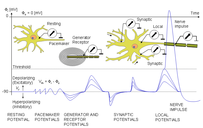
The axon has two states: resting and active. When the neurone is inactive the transmembrane potential is called the resting potential (nominally -70mV). When the nerve becomes active, it sends trains of short electrical pulses, called action potentials, to the end of the axon. Each action potential lasts ~1 millisecond during which the transmembrane potential is reversed, and these impulses are conducted - travel - along the axon at high speed (up to 120 m/sec), related to the diameter of the axon. Recording of action potentials can be done in two ways: action potentials in single axons requires microelectrodes that penetrate the nerve cell membrane, and second, the compound action potential can be recorded using external electrodes applied to a nerve containing many axons. The first method allows the mechanisms that generate action potentials to be studied; the compound action potential is useful to identify the different types of axons, and to study the condition of human nerves. |
|
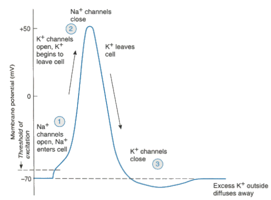
Action Potentials Action potentials are generated when voltage-gated sodium channels open as a result of the passage of local electrical currents across the membrane. These local currents may occur at the site of
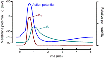
Whenever a depolarization of the axon is sufficient to reach a threshold (when the resting potential drops to around - 50 to -55 mv), voltage-sensitive sodium channels in the membrane open. As a consequence some Na+ ions move down both electrical and voltage gradients towards the Na+ equilibrium potential (about +40 to +60 mV) and cause the membrane potential to reverse for less than a millisecond. The All or Nothing Law (which applies to single axons) states that an electrical stimulus of a particular size to an axon either produces an action potential or it does not. The Threshold stimulus is the stimulus size (mV or mA) that just initiates an action potential A stimulus that is insufficient to initiate an action potential is known as a Subthreshold Stimulus A stimulus greater than the threshold stimulus is called a Suprathreshold stimulus: the action potential is no different from that induced by a threshold stimulus and the swing of membrane potential is constant in size |
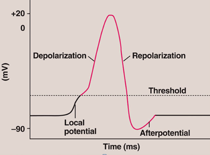 * *
This diagram shows the electrical changes during the action potential, elicited in response to a local potential reaching threshold. 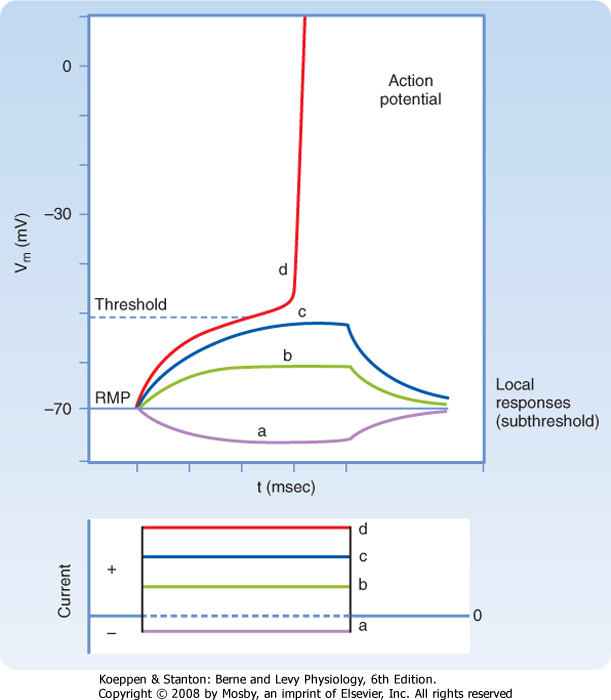 * *
|
Strength-Duration Curve and Refractory Period Top |
|
|
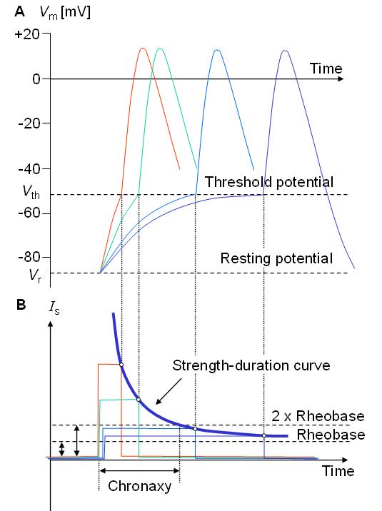
Strength-Duration Curve There is relationship between the strength of the stimulation current required to produce an action potential in a single axon, and the duration of that current. At longer the durations of stimulating current, less current is require to elicit an action potential. The strength-duration curve plots the relationship between these two variables. The hyperbolic shape of the graph shows the amount of current and its duration required to reach threshold. High currents require a shorter time to reach threshold. Lower currents require more time to reach threshold.
|
|
|
Refractory Period Following an action potential (AP) there is a short period of time (~1 msec) during which the nerve is refractory: i.e. it is irresponsive or less responsive to electrical stimuli. This is the time at which the voltage gated sodium channels are returning to their normal conformation (the channels are 'inactivated'). |
The Absolute Refractory Period is the period after the AP during which the nerve cannot be re-excited by electrical stimulation, no matter how large the stimulus. This is because Na+ channels are totally inactivated. In the Relative Refractory Period, a second AP can be initiated, but only by using larger stimulating currents. This is because some Na+ channels are still inactivated, while others are activated. |