Chapter 1 : The Cells of the CNS

Brain: Contents Page

Physiology of Nerve Cells : The Compound Action Potential

biologymad.com
Openi

Myelin and the Speed of Conduction of the Nerve Impulse

Schwann Cells and Myelination

In the peripheral nervous system, Schwann cells surround the axons and form a myelin sheath..

If axons are damaged, Schwann cells may also have phagocytotic activity and clear cellular debris that allows for regrowth of peripheral neurons down endoneurial tubes.

Schwann cells are also involved in the support of unmyelinated nerve axons. In this situation, no myelin sheath is formed, but several axons are surrounded by the Schwann cell, forming bundles of unmyelinated axons known as Remak Bundles.

In peripheral neuropathies, some Schwann cells may die and give rise to a pattern of segmental demyelination. This reduces the conduction velocities of the affected axons.


*

Oligodendrocytes

Oligodendrocytes are cells that coat axons in the CNS with their cell membrane forming a myelin sheath, that insulates the axon and is essential for fast transmission of nerve impulses. Defects in this system occur in demyelating diseases such as Multiple Sclerosis.

In contrast to the myelin of the peripheral nervous system, one oligodendrocyte can provide a myelin sheath for several axons.

Conduction of the Nerve Impulse

The area of the axon which is depolarised during the action potential sets up local currents: positve charges flow towards adjacent negative areas and depolarise the adjacent membrane. As a consequence the action potential is conducted along the axon.

*

Saltatory Conduction

Myelinated axons are surrounded by Schwann cells which provide insulation, and the voltage gated sodium channels are concentrated at the Nodes of Ranvier. This arrangement allows the currents flowing during the action potential to depolarise the next node of Ranvier, so the action potential jumps from one node to the next, a process called Saltatory Conduction (see opposite).

*


Top

Mammalian Nerves and the Compound Action Potential

Mammalian nerves consist of many axons running in parallel with each other. Larger diameter axons conduct action potentials faster than smaller fibres, and when electrical stimuli are applied, the voltage necessary to initiate an action potential in large axons is smaller than for smaller ones. When the stimulation voltage is increased, the first axons to generate and action potential are the large fast conducting axons. As the voltage is increased, smaller axons begin to generate an action potential, and these potentials are conducted relatively slowly. If electrodes are attached to the end of the nerve distant to the stimulating electrodes, the first potentials recorded are generated within the fast conducting large axons, and it takes more time for the action potentials of smaller axons to arrive at the recording site.

Nerves

Axons are cylinders of cytoplasm surrounded by the nerve cell membrane. They start at the axon hillock (the initial segment - about 25 micrometres long) can divide many times and each branch ends in a synaptic bouton. Axons can be up to 20 micrometres in diameter, or as little as 0.2 micrometres in diameter. The larger axons in mammals are myelinated - they have a myelin sheath produced by Schwann cells in the periphery and Oligodendrocytes in the CNS.

Axons can be short or very long - about a metre in the corticospinal tract axons that pass from the motor cortex to the lumbo-sacral cord. They are specialised processes which conduct electrical signals (action potentials) very rapidly to the nerve terminals.

In peripheral nerves, the axons are formed into bundles. The axons are surrounded by Endoneurium, and the bundle or fasciculus is surrounded by the perineurium. The bundles are gathered together inside a peripheral nerve, which has a fibrous sheath called the Epineurium. The whole structure also contains blood vessels to supply the necessary nutrients and oxygen.

Some nerves contain only sensory axons, others only motor axons, while others are mixed nerves with sensory and motor fibres.

*

Transverse section of a spinal nerve.

The Compound Action Potential

Large diameter axons conduct action potentials faster than smaller ones, and the process is speeded up further by the presence of myelin.

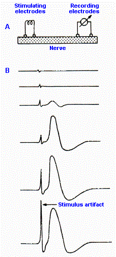
When electrical stimuli are applied (in A), subthreshold voltages do not elicit an action potential, but as the stimulus strength is increase, it is possible to record action potentials further down the nerve.

The conduction velocity can be calculated knowing the distance between stimulating and recording electrodes, and the time taken for conduction along the axons.

As the stimulus is increased all the largest fibres in the nerve are activated and cause a large peak of potential caused by the action potentials in all the fastest axons. This peak is known as the A-alpha wave.


*

Compound Action Potential

The top trace in the diagram opposite shows the A-alpha only peak over a longer time course.

The stimulus necessary to initiate an action potential in small axons is larger than for larger diameter axons.

As the stimulus is increased, smaller axons begin to generate their action potential, and these potentials are more slowly. Examples are the A-beta, A-gamma and A-delta potentials; it takes more time for the action potentials of smaller axons to arrive at the recording site.

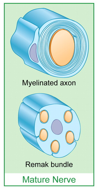
All of the A waves are due to action potentials in myelinated axons: A alpha axons have a lot lf myelin, whereas A-delta axons are finely myelinated.

Finally at very high stimulus strengths the unmyelinated C-fibres are activated, and it can be seen that the conduct very slowly.

Conduction Velocity

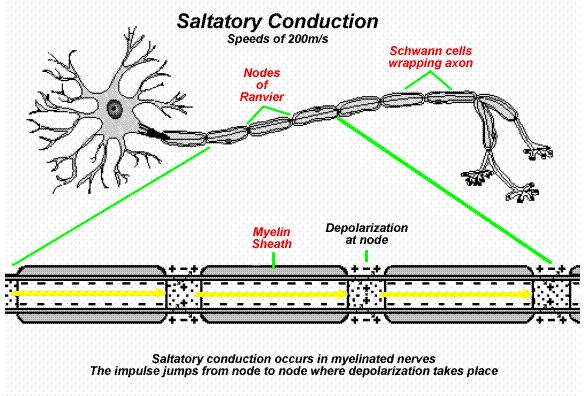
Axons conduct APs at different velocities, and the fastest use a process called Saltatory Conduction.

Large myelinated axons conduct rapidly (100+ m/sec).  The high speed is due largely to the myelin: 
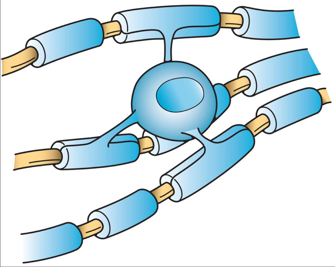
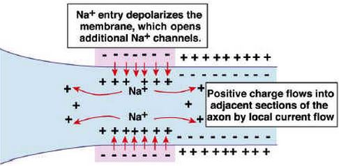
the AP jumps from one node of Ranvier to the next because currents follow pathways of least resistance, and myelin (rolled fatty cell membrane) has a high resistance

A fibres conduct at high speed A-alpha conducts more rapidly than A-beta and A-delta (because of the lower degree of myelination of the slower conducting axons). See Saltatory Conduction (below).

C fibres are unmyelinated and conduct very slowly (1 m/s)

*

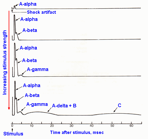
The thresholds of large axons are lower than the thresholds of small diameter axons. The diagram shows the effects of increasing stimulus strength, as a result of which more and more small axons become active. Smaller axons conduct more slowly and this is the basis of identifying A-beta (large myelinated), A-gamma and A-delta fibres (finely myelinated), and C (unmyelinated) axons.

For mammalian myelinated axons the conduction velocity (m/sec) is approximated 6x the diameter of the axon.


Top


Classification of Axons into different Types, based on Axonal Diameter and Conduction Velocity

Classification of axons (according to conduction velocity and diameter).

Relationship between diameter of axons and their conduction velocity.

Erlanger and Gasser classified motor axons into two groups of A fibres.
A fibres Diameter Conduction Velocity
A-alpha 13-20 micrometres 80-120 m/sec
A-gamma 5-8 micrometres 4-24 m/sec

Sensory Axons were classified using a different system of Fibre Types:

Fibre Type

Erlanger -Gasser Classification

Diameter (micrometres) Conduction Velocity (m/sec)
Ia and Ib A-alpha 13-20 80-120
II A-beta 6-12 31-80
III A-delta (B) 1-5 3-30 (B: 3-15)
IV C <1 unmyelinated <2

Erlanger and Gasser classified the preganglionic sympathetic fibres as Group B, and the post-ganglionic ones as Group C.

Different groups of nerve fibres in the Musculo-Skeletal System and in Skin have different functions:

Large myelinated motor axons in Muscle nerves consist of::

  • A alpha: motoneurones to skeletal muscle
  • A gamma: motoneurones innervating muscle spindles
Unmyelinated motor axons in muscle nerves belong to the autonomic nervous system and supply blood vessels

Large myelinated sensory axons in muscle nerves sense joint position and are involved in thecoordination of movement:

  • Ia - muscle spindles
  • Ib - Golgi Tendon Organs
  • Small myelinated and unmyelinated fibres in muscle nerves are responsible for pain and deep pressure sensations

    • Group III and IV fibres

    Different groups of sensory axons in cutaneous nerves have the following functions:

    • A-beta : touch
    • A-delta : coarse touch and fast pain
    • C fibres : Pain and Temperature

    http://usmanscience.com/
    michaeldmann.net
    michaeldmann.net
    docstoc

    Brain: Contents Page

    Top