Chapter 1 : The Cells of the CNS

Brain: Contents Page

Physiology of Nerve Cells: Axonal Transport

Axoplasmic Transport

Axoplasmic Transport is the transport of chemicals, vesicles and cell organelles along the interior of the axon

  • from cell body to nerve terminals (anterograde axonal transport), and
  • from nerve terminals to the cell body (retrograde axonal transport).

The cytoplasm of axons contains neurotubules which are the transport system along which packages containing essential materials are transported through the axoplasm.

RNA is contained in the Nissl substance of neuronal cell bodies, and proteins and peptides are synthesised there. In addition to the enzymes, neurotransmitters and structural proteins, there is a rate of turnover of cell organelles, all of which need to be replaced regularly, and anterograde axonal transport is essential for the delivery of these to the axon terminals.

The proteins kinesin and dynein are involved in the transport process along neurotubules, and the speed of transport is sometimes divided into two types: fast and slow.

'Fast axonal transport' is extremely slow compared with the speed of the action potential - around 100 mm/day. Vesicles are transported by the fast mechanism.

'Slow axonal transport' is much slower - around 1 mm/day and neurofilaments appear to use this slow mechanism..

*

In orthograde (anterograde) axonal transport packages of materials are transported within the axon from the cell body to the axon terminals. These packages contain a mix of components required for each function, within a vesicle.

Reterograde axonal transport is the process whereby substances are carried from the terminal boutons to the cell body. Some vesicular material is recycled and retruned to the cell soma. The contents of the retrogradely transported vesicles include chemical messages released by the post-synaptic cell.

The Cytoskeleton

The cytoplasm of neurones also contains neurofilaments and neurotubules.

Neurofilaments are synthesised in the cell body during the course of regeneration. They are needed to extend the axon and increase the diameter of the regenerated axon terminals.

Neurofilaments are a major component of the neuronal cytoskeleton, and provide structural support for the dendrites and the axon and regulate axon diameter. 

Glia do not contain neurofilaments.


*

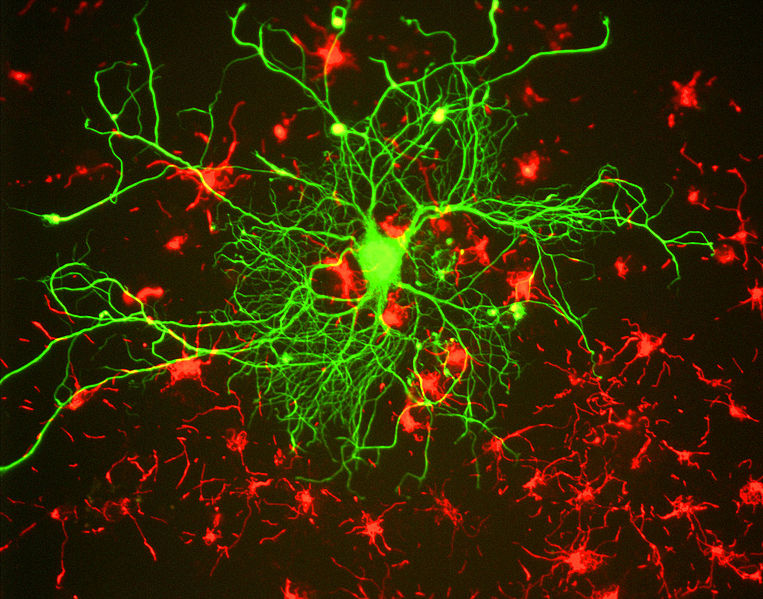
Rat brain cells grown in tissue culture and stained, in green, with an antibody to neurofilament subunit NF-L, which reveals a large neuron.

www.studyblue.com
Gery Shaw via Wikipedia Commons

Top

Anterograde (Orthograde) and Retrograde Axoplasmic Transport

Anterograde Axoplasmic Transport

Anterograde (Orthograde) Axonal Transport is the mechanism whereby packets of chemicals and organelles are transported within the axoplasm between the cell body and the synaptic boutons.

The chemicals necessary for the nutrition and maintenance of the nerve terminal are mainly manufactured in the cell body and their transport to the synaptic bouton is achieved by packages passing down the neurotubules of axons using the dynein/kinesin system of proteins.

The supply of enzymes and peptide transmitters necessary for some synaptic functions depends upon their synthesis in the neuronal cell body, and passage to the nerve terminals using axoplasmic transport.

Packages travel in vesicles, and dense-cored vesicles carrying peptides can be found along the length of the axoplasm.

Retrograde Axonal Transport.

Retrograde axonal transport carries chemical packages back to the cell body from the axon and nerve terminal. These chemical messages relate to the integrity and effectiveness of synaptic contacts.

At the pre-synaptic membrane clathrin-coated vesicles are believed to be involved in endocytosis - the formation of vesicles that sample the extracellular fluid within the synapse.

Growth factors such as neurotrophins are believed to be passed from the post-synaptic cell into the presynaptic terminals, and are transported to the neuronal cell body.

This process is of importance for the neurone which needs to know whether it is in functional contact with the post-synaptic cell. This retrograde transport of materials lets the neurone know about the state of the contact with the post-synaptic cell.


Top

Axoplasmic Transport in Disorders of Peripheral Nerves

Axonal Transport and Nerve Regeneration

After nerve section, clearly the retrograde transfer of these chemical signals stops, and it is the lack of the normal signal that causes the processes that follow nerve transection: chromatolysis, and protein synthesis.

Synthesis of structural proteins and transport of the essential materials for the formation of the growth cone and the extension of axons is an essential part of the process of nerve regeneration.

In the process some neurones undergo apoptosis - programmed cell death, so the number of neurones innervating a muscle is usually reduced; this is one factor in leading to the large motor units after regeneration of motoneurones.

When a regenerating axon again establishes contact with the post-synaptic cell (say skeletal muscle) retrograde chemical signals passed along the axon confirm that the synapses are again functional.

Tetanus: the Infection.

The pathology of the disease tetanus, caused by the release of tetanospasmin (also called tetanus toxin) by the anaerobic bacterium Clostridium tetani within the body depends upon the retrograde transport of tetanospasmin into the nervous system.

The toxin is transported retrogradely along motoneurone axons to the motoneurone cell body.

It then crosses synapses into interneurones that synapse on motoneurones and block the release of inhibitory transmitters, such as GABA and Glycine. This lack of inhbitory influence on motoneurones causes them to fire action potentials continuously and generate the characteristic severe muscle spasms that can be fatal.

The muscles first affected are those with short nerves, so before the generalised convulsions occur, muscles close to their motoneurone cell bodies give the early clinical signs of the disease.

Risus sardonicus is an early sign in which the face has a fixed smile and the patient has difficulty opening his mouth (lockjaw). These are due to overactivity in the facial (VII) and trigeminal (V) nerves, related to the loss of inhibitory influences of Glycine and GABA on V and VII nerve motoneurones.


Top

Axoplasmic Transport in Neurodegenerative Diseases.

Some of the pathological changes in neurones of patients with neurodegenerative diseases show signs of a disruption of axoplasmic transport by the microtubules.

These diseases include Alzheimer's disease, Parkinson's disease and Motoneurone Disease

One characteristic change in neurones in Alzheimer's disease is the presence of neurofibrillary tangles, which are are collections of twisted protein threads present within nerve cells.

Normal microtubules transport vesicles down the axoplasm, and are stabilised by the presence of a protein called tau.

In Alzheimer's disease an abnormally large number of additional phosphate molecules are found to be attached to tau, and allows the structure of microtubules to disintegrate, and at the same time hyperphosphorylated tau joins with other filaments to form a sort of mesh. This leads to a total disruption of the internal communication within the nerve cell, and a failure of axonal transport in both directions.


The interference with internal transport systems of neurones in these disorders results in an inability of the cell body and axon terminals to communicate with each other, so the chemicals normally processed at nerve endings are no longer available. Synaptic transmission is therefore affected and neurones fail to communicate with each other. In addition there is evidence of an accumulation of vesicles and filaments along the axons.

A consequence is that the neurones involved begin to die, and affected targets of the cortex begin to shrink and , as a result the brain atrophies. In severe Alzheimer's disease shrinkage of the brain is widespread. In fact disorders of axonal transport are probably involved in the pathogenesis of many neurodegenerative diseases.

Another pathological change in the brains of patients with Alzheirmer's Disease is the presence of insoluble deposits of beta amyloid. These pathological changes within the nervous system in patients with Alzheimer's disease are the result of abnormal prcessing a a normal protein in the cell membrane called amyloid protein precursor (APP). Abnormal processing produces peptide fragments that are toxic to nerve cells.

Top


Brain: Contents Page

Top

© HumanPhysiology.Academy 2014-2015