|
Membrane Capacitance: Physics The nerve cell membrane has a capacitance that depends on the properties of the lipid bilayer, and is fairly constant. The properties are evident when subthreshold depolarising currents and hyperpolarising currents are passed across the cell membrane. Many diagrams show the membrane potential as a series of negative charges lined up on the inside of the membrane, and positive charges on its outside, just as charge builds up on each plate of a capacitor. When a subthreshold current enters the cell, the voltage change reaches a peak and then declines gradually due to the capacitance of the membrane; the time it takes depends on the membrane time constant, τ. The time constant equals the product of membrane resistance and membrance capacitance (τ = RC). Once the current flow that has caused a new transmembrane potenial to be reached, it will return to its former state along a time course that depends on the membrane time constant. |
Local potentials: Electrotonic Spread When small amounts of current enter or leave the cell, they cause small depolarisations or hyperpolarisations, and the voltage change spreads around the focus of current flow. For example in a dendrite, local current flow at one synapse will cause depolarisation not just beneath the synapse but the potential change spreads locally and declines exponentially along the length of the dendrite, depending on the length constant. If another synapse, at a small distance from the first, becomes active, the potential changes from both can summate. These effects of depolarisations and hyperpolarisation at different (excitatory and inhibitory) synapses will summate algebraically and influence the membrane potential at some distance from their origin (depending on the length constant of the membrane). When the axon hillock becomes sufficiently depolarised, voltage-gated sodium channels at that site open when a threshold potential is reached, and the neurone fires one or more action potentials (see below). The greater the depolarisation at the axon hillock, the higher the frequency of the action potentials. |
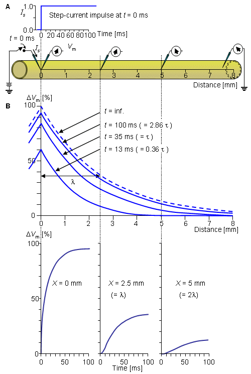
When small amounts of current flow through the membrane (i.e. currents insufficient to reach the threshold of an action potential) the potential changes both in time (depending on the time constant) and space (depending on the length constant). The length constant (λ), corresponds to the distance where a graded potential has decreased to 37% of its original amplitude. Individual cells have different length constants because the length constant is dependent upon the membrane resistance and the axial resistance; axial resistance is the resistance of the cytoplasm contained within the dendrite or axon, and depends on the diameter of the structure. Large processes (dendrites or axons) have longer length constants than thinner ones, so small graded potentials can travel over longer distances in the larger structures before they die out. In the diagram opposite a square wave depolarisation is applied theough the stimulating electrode in panel A. In Panel B, the potential change recorded at different distances form the stimulating site is measured, and the exponential decline in potential recoded at different distances fromm the stimulating site is due to the space constant of the membrane. In the bottom Panel (opposite), the time taken to achieve a new potential can be seen. Instead of the membrane potential changing instantly, it takes time to achieve a new steaty level, due to the time constant of the membrane. The three recordings show the changes at different distances from the stimulating site. These changes are due to the resistance and capacitance of the membrane, and an electrical model of the membrane (known as the cable properties) contains resistors and capacitors as shown below.
Cable properties account for the differences in electrical properties between small and large diameter axons. |
When a square wave current is injected into the axon (A). the trans-membrane potential at different distances from the electrode can be seen to change according to the distance from the site of current injection (top), and have a different time course at each site (bottom). These changes can be ttributed to the capacitance of and resitance of the cell membrane, a model of which is shown opposite. |

