Close

Glutamatergic Neurotransmission

Glutamate in Pre-Synaptic Endings

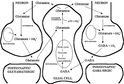
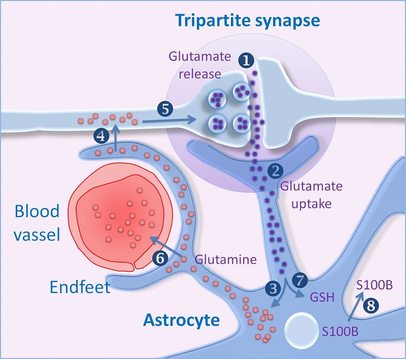
The amino acid Glutamate is synthesised from glutamine and stored within vesicles in synaptic boutons.

After its release into the synapse glutamate is transported into astrocytes which convert it into glutamine, which is recycled by the nerve endings. This is the basis of the Glutamine Cycle.


Glutamate Synthesis

Neurons synthesise glutamate from glutamine.

Glutamate is synthesised within glutamatergic nerve endings, but after release, it needs to be removed from the synapse quickly because accumulation of extracellular glutamate is associated with neuronal toxicity (see below).

The glutamate-glutamine cycle

The glutamate-glutamine cycle is recognised as an important metabolic pathway for mopping up excess glutamate from the synapse.

It is a mechanism whereby glutamate released at synapses can transported into adjacent astrocytes (and neurones) using a family of transporters - the Excitatory Amino Acid Transporters (EAAT) - and converted to glutamine within the astrocyte.

This glutamine can be recycled and passed back into the nerve endings for the synthesis of glutamate. Excess glutamine is transported away from the brain in the blood stream.

GABA and Glutamine

A similar process occurs for GABA (right).

Glutamate Transport into Synaptic Vesicles

After it is synthesised in the nerve endings, glutamate is transported into synaptic vesicles. Glutamate concentration in the vesicle increases to many times that of the cytoplasm.

The high concentration within the vesicle is produced by an ATP-dependent proton transport system that lowers the pH of the synaptic vesicle and carries the negatively charged glutamate ions into the vesicle.

The transporter involved is a family of proteins called the Vesicular Glutamate Transporters (VGLUT).


*

*
www.bioscience.org
www.intechopen.com

Post-synaptic Receptors for Glutamate

There are four types of Glutamate Receptors on neurones: The AMPA and NMDA receptors are of particular interest.

The AMPA receptor is responsible for the normal EPSP generated by glutamatergic neurones

The NMDA receptor can have no effect or produce very strong EPSPs, which it does only when the membrane is already depolarised; this changeover is due to Magnesium ions being displaced from their binding site.


AMPA Receptors (AMPARs)   Top

AMPA receptors mediate fast synaptic transmission. The name is derived from the ability of a synthetic glutamate analog, AMPA to activate the receptor.  The membrane becomes more permeable to sodium and potassium, which move across the membrane according to their electrochemical equilibrium potentials, i.e. small numbers of sodium ions move into the cell and potassium moves out.

AMPA receptors (AMPARs) induce small depolarisations in the post-synaptic membrane (EPSPs). When two glutamate molecules bind to an AMPA receptor, the protein channel undergoes a conformational change that allows sodium and potassium ions to move along their electrochemical gradients. Sodium ions move into the cell and potassium ions move out simultaneously, so the voltage change is approximately half way between the equilibrium potentials of both ions. The result is a depolarision of a few millivolts.

NMDA receptors (NMDARs)   Top

N-methyl-D-aspartate is a pharmacological agonist of one type of glutamate receptor, which is named NMDARs after this chemical.

The NMDA receptor is ligand-gated AND voltage-gated: it binds glutamate and glycine, but the size of the current flowing into the cell depends crucially on the transmembrane potential.

The dependence on the ongoing membrane potential is dependent on the fact that a magnesium ion blocks the entry of sodium and calcium through the channel at the normal resting potential. Sometimes this is known as open channel block.

However, when the membrane is depolarised, e.g. by the action of glutamate on AMPA receptors, the magnesium ion dissociates from the channel protein, and allows the influx of sodium and calcium.

When the membrane potential returns to its normal level, magnesium again binds to the channel and blocks entry of sodium and calium ions, even when glutamate is bound to the receptor.

NMDA antagonists include Ketamine, Pheylcyclidine and Nitrous Oxide (laughing gas).

*

The NMDA receptor is a complex ion channel that is ligand gated and voltage gated. The binding of magnesium ions at the normal resting potential prevents entry of sodium and calcium, even if glutamate is bound to the receptor. When the membrane is sufficiently depolarised, Mg2+ is released from its binding site and strong currents flow through the pore.

Note that zinc, glycine, polyamines and PCP also interact with the receptor. PCP (phencyclidine) is an NMDA receptor anatagonist.

Within the cerebral cortex, repeated activation of NMDA receptors can modify the shape of dendritic spine synapses, and can alter the strength of the synaptic event. This is believed to play an important role in Long Term Potentiation (LTP), a phenomenon thought to be part of the process of neural learning and the laying down of memories.

One consequence of calcium entry to dendritic spines through NMDARs is the activation of (a) calmodulin kinase which plays a part in the induction of new AMPA receptors, and (b) the production of a retrograde messenger, possibly nitic oxide (NO), that can modulate release of transmitters by the presynaptic terminal.

*

Metabotropic and Kainate Receptors   Top

The metabotropic glutamate receptors, or mGluRs, bind with glutamate, but instead of causing ion movements across the cell membrane, they activate a biochemical cascade that makes modifications to other proteins, including ion channels. These changes can lead to changes in synaptic strength and excitability, including the modulation of post-synaptic responses and presynaptic inhibition.

Kainate receptors produce small slow depolarisations of post-synaptic neurones.


graulab.tamu.edu
www.frca.co.uk


Glutamate Toxicity   Top

When activated, the NMDA receptor allows calcium ions to enter the cell if the cell membrane is already depolarised. Normally this entry of calcium ions causes a powerful depolarisation of the neurone, but these calicum ions also activate enzymes, such as some G-proteins.

Activation of G-proteins may increase the size of dendritic spine synapses, and alter the strength of the synaptic event. This change in synaptic strength is thought to be the mechanism underlying learning associated with repetitive activity, and the laying down of memories.

Excitotoxicity is the pathological process by which neuronal degeneration or death can occur by excessive stimulation by neurotransmitters such as glutamate.

Pathologically high levels of glutamate cause toxicity by allowing excessive quantitites of calcium ions to enter the neurone. These high levels can activate certain intracellular enzymes concerned with the degradation of phospholipids, proteins and nucleic acids, which, in turn, damage intracellular structures such as the cytoskeleton, membranes including that of intracellular organelles, and nucleic acids.

Excitotoxicity may be involved in neuropathology including conditions such as a range of neurodegenerative diseases, stroke, traumatic brain and spinal cord injuries. Hypoglycemia and status epilepticus may cause excessive glutamate concentrations around neurons.

Top Close

free hit counter

© HumanPhysiology.Academy 2014-2015