Close

Types of Inhibitory Mechanisms

Post-synaptic Inhibition of a neurone occurs because of the hyperpolarisation produced by the action of an inhibitory neurotransmitter on a neurone.

In Pre-synaptic Inhibition the amount of excitatory transmitter released by a specific synaptic bouton is reduced, and occurs at axo-axonic synapses.


Top Close

Pre-Synaptic Inhibition

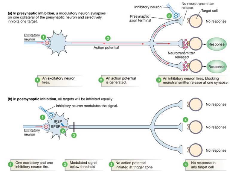
Presynaptic Inhibition is a mechanism by which the amount of neurotransmitter released by an individual synapse can be reduced, resulting of less excitation of the post-synaptic neurone.

When this occurs the 'inhibition' is actually due to less excitatory input.

What's more the 'inhibition' is targeted at specific types of input to a neurone, in contrast with the IPSP, which acts post-synapticially, and inhibits all activity in the neurone.

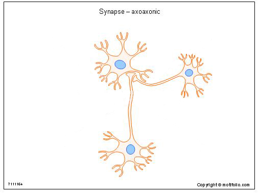
The anatomical basis of the process is the axo-axonic synapse, a diagram of which is shown opposite. The neurone on the right has its axon terminating on the synaptic bouton of the bottom neurone. These axo-axonic contacts are found in many parts of the CNS, including the dorsal horn of the spinal cord.

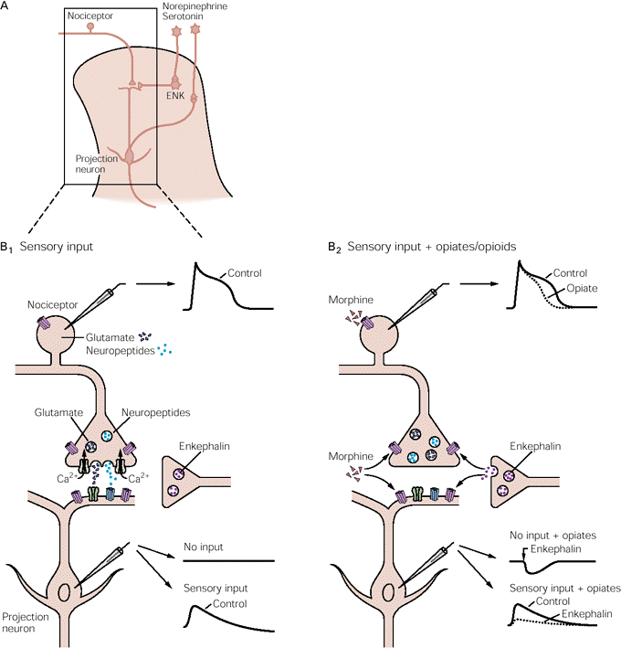
The physiological significance of the axo-axonic synapse is the existence of primary afferent depolarisation, and is of importance because of their involvement in the modulation of nociceptive messages.

*
Primary Afferent Depolaristion (PAD)

In the diagram opposite, synaptic bouton H forms an axo-axonic synapse with bouton F.

Activation of bouton H has no effect on post-synaptic neurone Y, but it does reduce the effects of stimulating axon F. This occurs because of primary afferent depolarisation (PAD) generated in bouton F.

PAD is the process of depolarisation of terminal F, which may seem surprising. It works because the calcium entry that occurs when an action potential arrives in F is reduced as a result of starting from a depolarised state. The calcium entry can also be reduced by reducing the duration of the calcium current, which characteristically has a small plateau.

So the release of transmitter by F can be reduced if, as a result of activating H, the amount of calcium entering F is reduced because the membrane starts from a depolarised state (i.e. the 'swing' in calcium potential in less), or because the duration of the calcium current is shorter.


*

PAD in the Dorsal Horn.

The diagram opposite shows one important site where primary afferent depolarisation has important effects in the pain pathway. PAD in nociceptive afferents is mediated by the endogenous opiate Enkephalin.

Left: activity in the nociceptor excites the post-synaption projection neurone using glutamate or neuropeptides. The action potential at the terminal has a plateau due to calcium entry, and this causes the release fo vesicles of neurotransmitter.

Right: the effects of the opiate include a reduction in the duration of the calcium current in the terminal produced by the release of enkephalin, which results in a reduction in glutamate release and an effective block in nociceptive transmission.


*

ib.cnea.gov.ar
Pearson and droualb.faculty_mjc_edu
www.motifolio.com

Top Close


Comparison of Pre- and Post-Synaptic Inhibitory Mechanisms   Top

*

This diagram shows that IPSPs affect the total activity of a neurone. In contrast, presynaptic inhibition of one of the neurone's terminals (top) affects only that individual synapse.
faculty.pasadena.edu

Close

© HumanPhysiology.Academy 2014-2015