Chapter 2 : The Spinal Cord

Brain: Contents Page

An Overview of the Central Nervous System : The Spinal Cord


Spinal Cord Gross Anatomy: Segmental Structure

A diagramattic explanation of anatomical terms such as rostral, caudal, superior, inferior, dorsal, ventral, coronal, sagittal, etc can be found here: Some Anatomical Terms

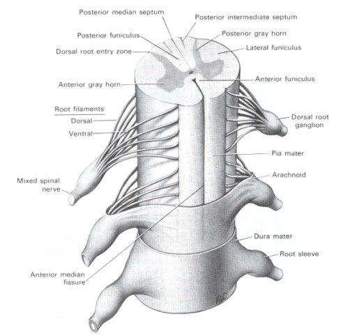
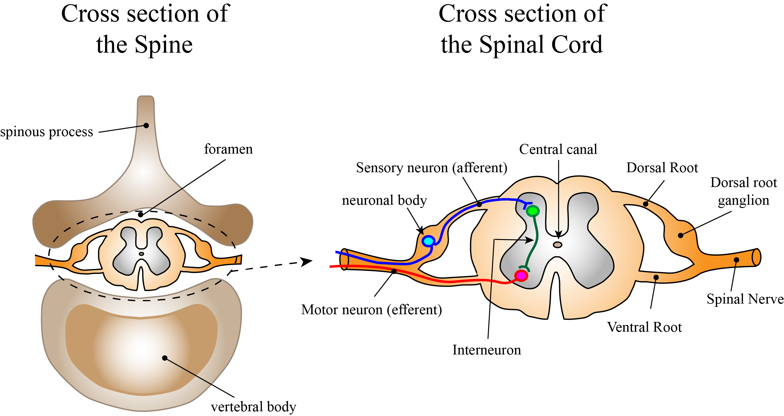
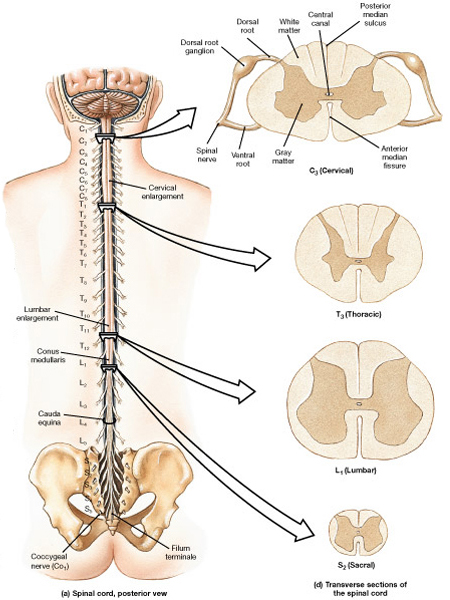
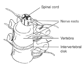
The spinal cord has a cylindrical segmented structure, each segment being derived from the somites of the embryo. A spinal nerve arises from each side of every segment and these segmental nerves leave the vertebral canal through the inter-vertebral foraminae.  

The spinal cord is protected by the vertebral column, to which the outermost of the meninges, the dura mater, is attached.

Movements of the vertebral column are buffered by the presence of cerebrospinal fluid within the subarachnoid space - so the spinal cord floats in a tube of fluid between the arachnoid and pia mater.

The spinal cord ends at the second lumbar segment of the vertebral column, below which the lower lumbar, sacral and coccygeal nerves travel through the subarachnoid space to reach the appropriate intervertebral foraminae where they leave the spinal canal.

These nerves attached to the bottom of the cord appear like a horese's tail and are calles the 'cauda equina'.


*

Spinal White Matter

The outside of the cord is white because it contains many parallel myelinated axons, some running the full length of the cord to the brainstem and beyond. It also contains axons that descend from the cerebral cortex (the cortico-spinal tract) and structures in the brainstem (reticulo-spinal tracts) that synapse on neurones in the grey matter

Dorsal Columns

The dorsal columns contain the axon collaterals (branches) of sensory neurones which synapse on neurones in the medulla. The information carried by these axons is concerned with touch and vibration.

Lateral Columns

The lateral columns contain axons of dorsal horn neurones that ascend to the thalamus and cerebellum, and descending axons originating from neuronal cell bodies in the cerebral cortex and structures in the brainstem.

Anterior Columns

A small number of cortico-spinal tract axons descend in the anterior columns of the cord.

More information about Tracts and their functions can be found in the section on Tracts in the CNS.

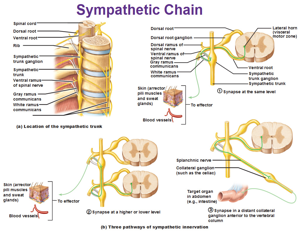
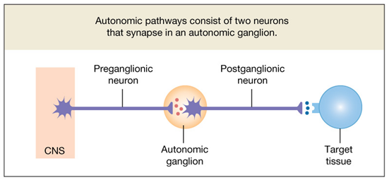
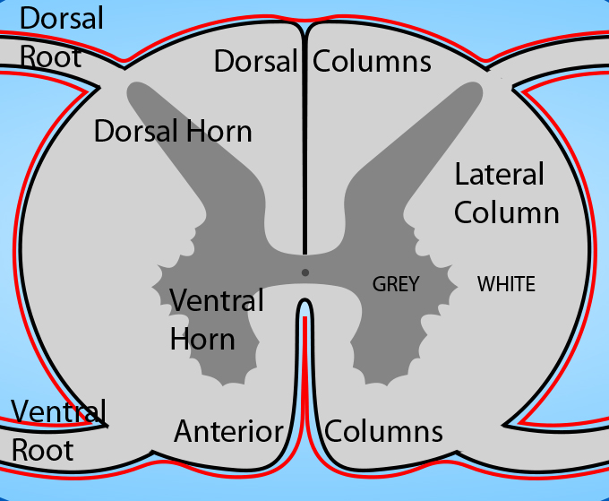
The red lines represent the pia mater.

Spinal Grey Matter

All segments of the cord have grey matter in an H-shaped arrangement around its central canal. It is divided into two horns : dorsal and ventral. In some segments there is a small additional horn- the lateral horn, adjacent to the central canal, that is important for autonomic functions.

The grey matter consists mainly of neuronal cell bodies and synapses, whereas the white matter consists of bundles of fibres, grouped together into tracts which project to specific destinations.

Vascular supply to the spinal cord.    Top

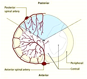
The arterial supply of blood to the spinal cord comes through the anterior spinal artery (which runs ventrally in the midline), and two other arteries that run longitudinally near the dorsal root entry zone.   These vessels get their blood from the vertebral arteries; other arterial vessels enter the cord along the dorsal and ventral roots, and anastomose with the other vessels.

Occlusion of arterial vessels supplying the cord causes damage or death to the neurones and axons within their territory, and the diagram below shows the distribution of these vessels. Note that the anterior artery supplies the ventral horn and the lateral and anterior columns.

Occlusion of the anterior spinal artery is characterized by loss of motor function and pain and temperature sensation below the level of occlusion but with preservation of touch sensation and proprioception.

The symptoms of anterior spinal artery occlusion can be explained by interruption of the corticospinal and anterolateral pathways and preservation of the dorsal column pathways. More details in the section on Tracts in the CNS.

*

http://www.laesieworks.com/spinal/SCIinfo01.html
http://bellspalsycranialnerves.wordpress.com/tag/spinal-cord/

Top


Spinal Cord Gross Anatomy: Cervical and Lumbar Enlargements

Each spinal nerve has two roots which contain axons with different functions.  Each dorsal root has a ganglion which contains the cell bodies of sensory neurones that transmit information into the spinal cord from skin, muscles, joints and viscera. These are often called afferent nerves because information is passed into the CNS.

The ventral root does not have a ganglion and contains the axons of motoneurones located in the ventral horn that exit the vertebral canal to connect with skeletal muscles, and control their force of contraction and length. These axons conduct information in an efferent direction, i.e. away from the CNS.

In certain segments of the cord neurones of the autonomic nervous system pass through the ventral roots to autonomic ganglia which regulate the activity of visceral organs and blood vessels. The cell bodies of these autonomic neurones are found in the lateral horn of the grey matter of the 1st thoracic to 2nd lumbar, and sacral segments 2-4 (see below).


Cervical and Lumbar Expansions of the Cord.   Top

The spinal cord is cylindrical, but it is wider in two areas- the cervical region (neck) and the lumbar region (lower back).  Cross sections of the spinal cord at different segments show thee differences, and some of the reasons for these expansions. 
The cervical expansion is partly due to the large motor innervation of the muscles of the upper limb.

Another reason is the large number of sensory axons that travel rostrally within the dorsal columns, and their numbers increase greatly in the cervical region because of the entry of large numbers of axons carrying sensory information from the hands and fingers. Similarly the lumbar expansion of the cord is due to the large number of efferent and afferent neurones carrying information to and from the lower limbs.

The left side of the diagram shows some of the differences between different cord segments. In particular notice the different in size of the cervical and lumbar expansions relative to the thoracic and sacral cord.

The dorsal columns contain the axon collaterals (branches) of sensory neurones which travel headwards and synapse on neurones in the medulla.

The axons from lower segments of the cord travel upwards near the midline. As we move up the cord, more afferent fibres enter the dorsal columns and they are added lateral to the existing ones. So the cervical segments the dorsal columns are much larger than the lumbar ones.

The ventral horn consists of the cell bodies of motoneurones, and the size of the ventral horn in the lumbar region is greater than in the thoracic region because of the large motor innervation of muscles in the lower limb. Similarly in the cervical cord, many motoneurones are used to control the upper limbs, hand and fingers, so the cervical ventral horn is large.

*

Spinal Cord Injuries   Top

Spinal cord injuries are commonly associated with fractures and displacement of the vertebral column.  The injury may cut through axons that carry messages between the brain and the motoneurones, or between the sensory nerves and the cerebral cortex.  In cases where the cord is totally transacted by the injury there is a  total lack of sensory and motor function below the level of injury.

Severe damage to the spinal cord can also occur as a result of occlusion of the anterior spinal artery, which if prolonged, can lead to infarction of the ventral regions of the cord and neuronal death.

More Details on Spinal Cord Injury

*
Wikipedia
http://what-when-how.com/neuroscience/
http://droualb.faculty.mjc.edu/Lecture%20Notes/Unit%205/

Top


Autonomic Nervous System : Anatomy

The autonomic nervous system is a system of efferent nerves that connect the CNS with internal organs, but unlike the motoneurones that connect directly with skeletal muscles, the pathway between the spinal cord and a visceral organ is interrupted by a synapse within an autonomic ganglion.

The presence of the ganglion and synapse in the path of the nerve defines the pre- and post-ganglionic neurones of these pathways. The cell bodies of the preganglionic neurones are within the CNS, whereas those of the post-ganglionic neurones are within the ganglia.

The preganglionic neurones are generally finely myelinated, whereas the postganglionic neurones are unmyelinated. Preganglionic neurones use acetylcholine as their neurotransmitter within the autonomic ganglia.

*


*

Lateral Horn of the Spinal Cord

The lateral horn contains the preganglionic neurones of the autonomic nervous system; this area is sometimes given the name intermedio-lateral column because some neurones have their cell bodies in a more medial position.

However, a lateral horn is only present in certain segments of the cord, which are:

the first thoracic to second lumbar segments, and sacral segments 2-4 (in humans).

So the lateral horn is absent in the cervical cord and the lower lumbar cord.

The neurones of the lateral horn between the first thoracic and second lumbar segments are referred to as the sympathetic preganglionic neurones. The axons of these neurones synapse on post-ganglionic neurones in the sympathetic chain, - a chain of ganglia running alongside the vertebrae throughout the length of the vertebral column including the neck and pelvic regions. 


In addition some innervate the adrenal medulla and some specialised pre-vertebral ganglia in the abdomen, such as the coeliac or inferior mesenteric ganglia.

The neurones of the lateral horn of the sacral cord send axons to parasympathetic ganglia that are locate close to the internal organs of the pelvis, where they synapse of postganglionic neurones that innervate the pelvic urogenital or colo-rectal organs.

The parasympathetic system includes a cranial outflow that innervates the intrinsic muscles of the eye, the salivary glands and thoracic organs, and the upper 2/3rds of the gastrointestinal tract, as shown in the diagram opposite.

Greater coverage of the autonomic nervous system will occur in the section on autonomic physiology.

Myelination of preganglionic axons

Pre-ganglionic fibres have a fine degree of myelination and are therefore whiter than the grey, unmyelinated, post-ganglionic axons. 

The difference in colour is discernible in the white and grey rami communicantes which connect the sympathetic chain and the spinal nerves.

The myelinated preganglionic axons pass through the white rami comunicantes on their way to the sympathetic ganglia. Unmyelinated axons of postganglionic neurones pass through the gray rami communicantes to re-enter the spinal nerve and continue towards their target organs such as blood vessels.

*

classroom.sdmesa.edu
antranik.org
faculty.pasadena.edu

Top

Chapter 2 : The Spinal Cord

Brain: Contents Page

© HumanPhysiology.Academy 2014-2015