Sensory receptors can be classified by the tissue in which they are located, e.g. muscle, skin, joints, visceral organs, and can be further subdivided according to the morphology of their sensory endings, e.g. encapsulated endings or free nerve endings.
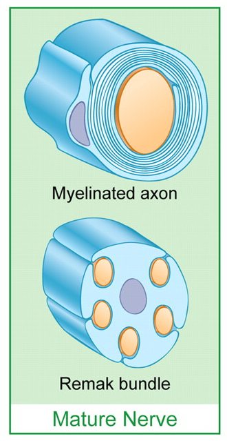
Neurones with cell bodies in the dorsal root ganglia have perikarya and axons of different diameters. These axons are often divided into groups depending on the degree of myelination: unmyelinated, finely myelinated and large myelinated. The axons with the largest diameters have the highest degree of myelination |
In these diagrams of peripheral nerves, several unmyelinated axons are invaginated into a Schwann Cell (forming a Remak Bundle) but myelin (i.e. multiple layers of cell membrane) is not formed around these axons. In myelinated axons, the Schwann cell wraps itself around the axon many times, and the multiple layes of cell membrane act as the fatty insulator called myelin. |
The Muscle Spindle The largest afferent axons in peripheral nerves innervate muscle spindles and tendons. These provide the nervous system with information about muscle length and tension, and contibute to kinaesthesia - the sense of position, particularly in a limb. Joint receptors are also involved in kinaesthesia. The Muscle Spindle is spindle shaped- i.e. has a bulge in its central region and is surrounded by a capsule; throughout its length it contains modified muscle fibres called intrafusal (meaning inside the spindle) fibres. The structure is orientated in a parallel direction with the much larger extrafusal fibres, and is attached to the endomysium that surrounds the extrafusal fibres. The muscles spindle has both afferent and efferent innervation. The largest afferent fibres are the Ia afferents arise from the a sensory ending that spirals around the central region of the intrafusal fibres- sometimes called the annulo-spiral ending. Other nerve fibres innervating the spindle include (a) a smaller diameter (group II) afferent and (b) some fibres that cause the intrafusal muscle fibres to contract. The latter are called gamma-efferent fibres and are finely myelinated. The Golgi Tendon Organ Golgi Tendon Organs arise from tendons and have large diameter axons that are slightly smaller than those of the muscle spindle. These afferents are sometimes called Ib afferents. |
Simple diagram of the Muscle Spindle.More details on Muscle Spindles 
Golgi Tendon Organ |
|
Joint Receptors |
|
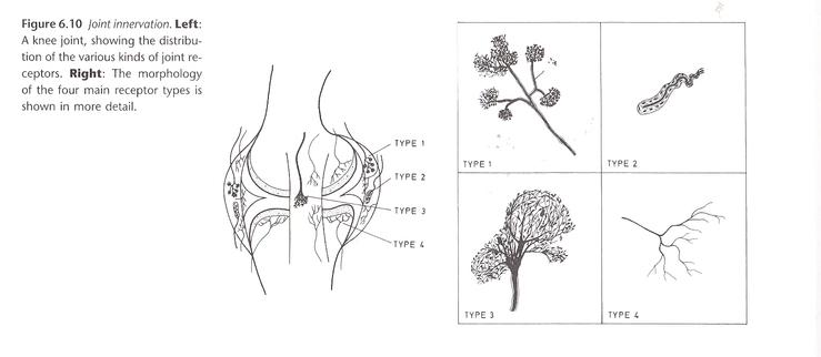 Joint receptors are found in the synovial membranes, joint capsules and ligaments, and respond to mechanical changes in joint position, and play an important role in kinaesthesia and proprioception. Four types of receptor endings are recognised in the synovial membranes and connective tissue/ligaments around joints. Ruffini endings akin to the Type II slowly adapting receptors found in the dermis are present in the connective tissues around joints. Paciniform corpuscles are akin to the Pacinian Corpuscles found in other tissues, but have less lamellae in the onion-like encapsulation of the ending. Golgi receptors and free nerve endings are also present around joints. These four receptors allow for the sensation of joint movement, joint position and the extremes of movement associated with pain. |
|
Sensory Receptors in the Skin Large axons also innervate the skin, and mediate the senses of Touch and Vibration. Merkel Cells exist in the basal layers of the epidermis and enclose the terminals of some large myelinated axons. Branches of one axon makes contact with many Merkel cells which sense indentation of the skin. These are the structures at the terminals of Slowly Adapting Type I receptors. Ruffini endings lie deeper in the dermis and have axon terminals wound round a central core within and encapsulated organ that is attached to the connective tissue in the dermis; these are associaed with Type II slowly adapting recpetor activity. Meissner's corpuscles are encapsulated end organs that also have myelinated axons, and are situated near the epidermal-dermal junction. They exhibit a complex array of nerves and lamellae within the structure of the ending. They are rapidly adapting receptors. Hair receptors have myelinated axons and innervate the hair follicle.
Vibration is sensed by Meissner's corpuscles and Pacinian corpuscles. Pacinian corpuscles lie in the deeper layers of the skin and large endings that might be likened to a small onion surrounding the terminal of a myelinated axon. They are very sensitive to high frequencies of vibration (~250Hz). Smaller versions of the Pacinian Corpuscle are found in many tissues and known as Paciniform Corpuscles, but the number of lamellae in the encapsulated ending is less. Meissner's Corpuscles are rapidly adapting receptors most sensitive to frequencies of ~30Hz. Hair follicles receive their own specialised innervation, and sense the presence of hair movement. Vibrissae (whiskers) have a dense innervation within their follicles. These skin receptors are not normally involved in initiating reflex activity. |
 * *
Note the difference between hairy skin and glabrous skin - the skin of the palm of the hand.  Pacinian Corpuscle Pacinian Corpuscle |
The free nerve endings of unmyelinated axons occur in almost all tissues, and the sensory ones generally mediate the senses of pain and temperature. The sensory receptors that respond to injurious stimuli are called nociceptors. They induce reflex activity in flexor muscles and in the autonomic nervous system. Nociceptors respond to injurious stimuli caused by
|
The mechanism by which nociceptors work involves membrane proteins called TRPV receptors . Transient Receptor Potential Vanilloid channels are a super- family of transient receptor potential (TRP) ion channels, that are selective for calcium and magnesium over sodium ions. Several members of the family are sensitive to elevated temperature, while others are sensitive to cold. The TRPV super-family of transducers show sensitivity to not only to heat and cold, but some respond to acid conditions, osmolality and capsaicin, a vanilloid molecule derived from hot red peppers. |