Chapter 2 : The Spinal Cord

Brain: Contents Page

Physiology of Reflexes

Reflex movements are movements initiated by sensory receptors, which, by having synaptic contacts within the spinal cord, are a basic level of regulation of muscles or glands. The Spinal Reflexes are the most basic of all reflexes, but other parts of the central nervous system also contain reflex pathways. Examples include the vestibulo-ocular reflexes, the vestibular reflexes affecting the body musculature in the control of balance, and the autonomic reflexes that regulate the diameter of the pupil or the degree of accommodation of the lens of the eye. But it is conventional to start in the spinal cord.


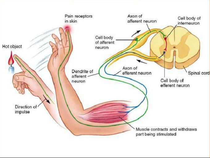
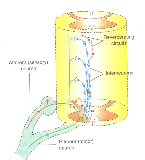
Spinal Reflexes   Top

The sense of perception is a function of the higher levels of the nervous system, so activity in pathways that exist in their entirety within the spinal cord are not accompanied by any sensation. Following spinal cord transection, patients have no knowledge of events occuring below the level of the lesion, and have no motor control over their muscles below that level. However neural pathways in the spinal cord below the lesion continue to function and these pathways are responsible for the reflex activities of the detached length of cord.

All reflexes have an afferent pathway that uses a specific type of sensory receptor, at least one synapse in the pathway, and an efferent pathway that connects to a muscle or gland. The site of the synapse is generally within the spinal cord, although the enteric nervous system also participates in reflex activity confined to the gut wall.

Synaptic transmission is unidirectional – from pre-synaptic ending on to post-synaptic membrane, because the neurotransmitter is released from the pre-synaptic ending and acts on the receptors in the post-synaptic membrane. Some reflexes are monosynaptic, such as the stretch reflex. Other reflex pathways are polysynaptic, and include interneurones that connect the sensory and effector neurones.

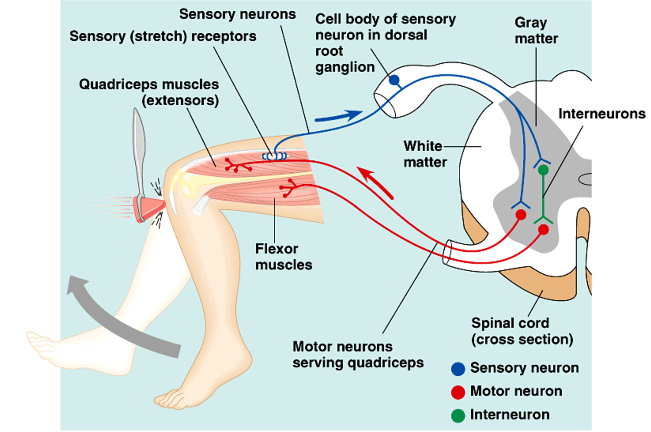
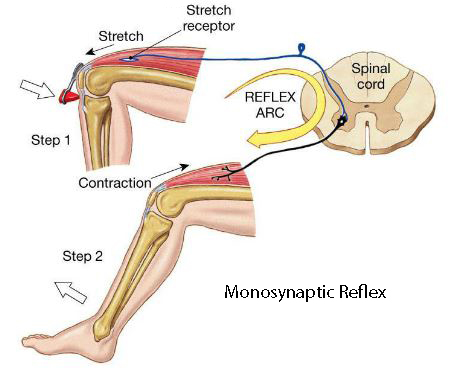
The simplest reflex arc is the monosynaptic (stretch) reflex.

The afferent fibres from the muscle spindles in a muscle enter the dorsal root and proceed to the ventral horn of the spinal cord. There they synapse on motoneurones that project back to the same muscle, or muscles in the same functional group.

This, the simplest of reflex pathways, is preserved following spinal transection, and is tested by clinicians who use a tendon hammer to apply a small stretch to the muscle.

This reflex is called the stretch reflex or knee jerk reflex (and sometimes the myotatic reflex), because it is initiated by stretching the muscle.

The reflex is an essential part of the motor control system in the intact nervous system, and allows a dynamic, fast feedback to occur from the active muscles.

*

The stretch reflex has a number of names:

  • Myotatic reflex
  • Tendon reflex
  • Tendon jerk

All have one thing in common, i.e. sudden stretch of muscle spindles in a muscle induced by the tap of a tendon hammer causes a reflex contraction of the stretched muscle using the monosynaptic reflex pathway.

It may also be called the

  • Knee Jerk – in the case of the knee
  • Ankle Jerk - in the case of the ankle
  • Elbow Jerk- in the case of the Elbow
  • Jaw Jerk - in the case of the Jaw.

The Tonic Stretch Reflex  Top

Muscle spindles respond to changes in length of muscles and the speed of those changes. They are also acutely sensitive to vibration, and produce reflex activity when vibrators are applied to muscles. These reflex responses consist of a tonic contraction of the muscle that is subjected to vibration, and can be maintained for substantial periods of time. This maintained reflex response is sometimes called the Tonic Stretch Reflex, and can be used by physiotherapists to help preserve muscle tone and simulate mild exercise after certain injuries.


Pathways of different reflexes

Monosynaptic Stretch reflex
Ib disynaptic reflex - Clasp Knife Reflex
Flexor Reflex
Crossed Extensor Reflex


Docstoc

Top

Physiological Characteristics of Spinal Reflexes

Segmental Arrangement of Spinal Cord

The spinal cord is arranged as a series of segments, and the motoneurones that control the largest muscle groups are spread over several segments.

The afferent axons that enter the dorsal roots divide into axon collaterals that synapse on neurones in the same and adjacent segments (see the diagram opposite).

Some of these afferent collaterals end directly on motoneurones

  • that innervate the same muscle or
  • muscles with a synergistic (similar) action

For these, there is only one synapse - between the muscle afferent and the motoneurones - hence the term monosynaptic reflex.

However other axon collaterals of Ia afferents from the same muscles spindles have different destinations:

  • Someare involved in initiating relaxation of antagonistic muscles during the reflex contraction.
  • Others make contact with neurones that project to higher levels of the nervous system, e.g. the cerebellum

Reciprocal Innervation

All afferent axons release an excitatory neurotransmitter - glutamate. However the relaxation of antagonistic muscles requires another mechanism, involving axon collaterals that excite an interneurone that in turn releases an inhibitory transmitter.

This mechanism allows antagonistic muscles to be relaxed during the contraction induced by the stretch reflex, and called reciprocal inhibition.

The inhibitory transmitter that hyperpolarises the motoneurones of the antagonistic muscle is glycine.

Diagram of the paths of afferent neurones entering the spinal cord in the dorsal root: the axons divide to form axon collaterals that synapse in the same or adjacent segments of the cord (Cajal).

*

The pathway of reciprocal inhibition.

Flexion Withdrawal Reflex: a polysynaptic reflex.

When injurious stimuli are applied to the skin, there is reflex withdrawal of the skin from the source of the injury. 

The stimuli that elicit this response are:

  • excessive force sufficient to penetrate or cause damage to the skin,
  • excessive heat that would denature proteins in the tissues are applied to the skin,
  • other physical or chemical stimuli that cause injury

Although pain is a normal accompaniment of this process, the pathway involved in this reflex response is entirely within the spinal cord, and the withdrawal responses occurs without the sensation of pain because the spinal cord is not directly involved in perception.

Withdrawal of a limb from a source of intense heat, pressure, force etc utilises the contraction of flexor muscles The reflex response is called the flexor withdrawal reflex and involves:

  • nociceptive afferents in skin
  • a polysynaptic pathway within the spinal cord
  • activation of flexor muscle groups that remove the injured part from the nociceptive stimulus.
*

Nociceptors have small diameter fibres with low conduction velocities (A-delta and C fibres). This is in contrast to the fast-conducting afferents in the stretch and clasp knife reflexes.

The flexor withdrawal reflex is sometimes called the 'inverse myotatic reflex'.

As the flexion reflex involves afferent activity in nociceptors, the reflex is elicited at the same time as the ascending pathways concerned with pain and nociception. Further consideration of the latter will be covered later.

Nociceptors, like all afferents, use glutamate as a primary transmitter. However other neurotransmitters are present in these small fibre afferents, and in particular, the presence of Substance P is significant, and this peptide and other synaptic mechanisms can have longer lasting effects that contribute to pain and the longer duration events of the flexor reflex.

When nociceptors elicit a flexor reflex, the polysynaptic pathway of the crossed extensor reflex excites extensor motoneurones on the opposite side of the cord at the same time as flexor muscles ar contracting..

*

Interneurones of the Flexion Withdrawal and Crossed Extensor Reflexes

It is not sensible for the reflex withdrawal of a limb to be short lasting - the limb would return to the site of the noxious stimulus. So there is a mechanism that prolongs the flexion, and this involves circuits of excitatory interneurones that re-excite each other. These circuits that involve re-excitation of interneurones and are responsible for maintained flexor activity are known as reverberating circuits.


These reverberating circuits maintain flexion for a period of time following a painful stimulus - so the foot is removed from the painful stimulus, and the flexion is maintained. These reverberating circuits are also involved in maintaining a state of excitation of extensor motoneurones, so as to prolong support for the body while the opposite limb is flexed.

*
thesalience.wordpress.com
Docstoc
Docstoc
Docstoc

Top

Summary: Differences between the Reflexes

Reflex pathways differ in their

  • sensory receptors,
  • afferent fibre types (A-alpha, A-delta, C),
  • number of synapses, and
  • type of motor response

In the Stretch Reflex:

  • the sensory receptor is the muscle spindle
  • fhe afferent fibres and large and rapidly conducting
  • the reflex is monosynaptic
  • the motor response is seen in the muscle that is stretched (and synergistic muscles).

In the Flexor Withdrawal Reflex:

  • the receptor is the nociceptor
  • the afferent fibres are small in diameter and slowly conducting
  • the central pathway is polysynaptic and involves reverberating circuits
  • the motor response is seen in the flexor muscles that remove the limb form the injurious stimulus

In the Crossed Extensor Reflex:

  • the sensory recpetors are nociceptors in the opposite limb
  • the central pathways involves many interneurones
  • the interneurones are involved in reverberating circuits that cross the cord
  • the motoneurones involved are extensor motoneurones that maintain support for the body while the opposite limb is flexed.

Top


Central Pattern Generators

Central pattern generators (CPGs) are neural networks that produce a rhythmic motor activity that does not involve sensory feedback. It is a pure motor act consisting or alternating or patterned activity in muscle groups generated by a neural network.

Examples include the scratch reflex (opposite), swallowing, respiratory movements, and possibly even walking or swimming.

In the simplest CRGs, rhythmic activity is generated by alternating trains of EPSPs and IPSPs within a spinal neural network.

The Scratch Reflex is a movement of the hindlimb of a spinal animal in response to a mild repetitive mechanical stimulus to the skin. The movement often consists of repeated alternating movements that attempt to remove the source of the stimulus.

This rhythmic response, which can be seen in domestic animals, is due to circuits of interneurones present within the spinal cord. These circuits involve many neurones within different spinal segments, and are given the name 'Central Pattern Generator'.

Top


Use of the Stretch Reflex in Clinics

Neurologists use the stretch reflex regularly to assess the strength of reflexes, which can be absent, present, normal or hyperactive.

In patients with Stroke and some other conditions, the activity of reflexes is increased and there is an absence of wasting of the muscles, which would only occur if a lower motoneurone lesion was present.

Loss of Reflex Responses

One cause of a loss of reflex responses is because a muscle becomes denervated. The characteristics of a lower motoneurone lesions are :

  • loss of voluntary movement
  • loss of electrical activity or movement on electrical stimulation of a motor nerve
  • loss of reflex activity
  • atrophy of the muscle

Causes of Lower Motoneurone Lesions

Occasionally the cause of a loss of reflex activity is due to lesions of the afferent limb of the reflex.


Top

Chapter 2 : The Spinal Cord

Brain: Contents Page

© HumanPhysiology.Academy 2014-2015