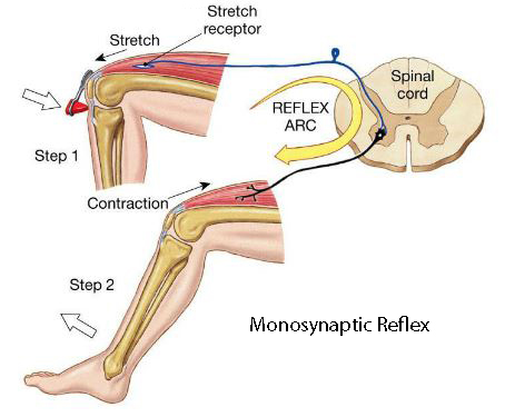
Monosynaptic Reflex The simplest reflex arc: the monosynaptic (stretch) reflex. On entering the spinal cord the sensory axons often divide to form axon collaterals. So all axons distribute their information to multiple groups of post-synaptic cells. These post-synaptic cells may be in the same segment or adjacent segments. For the largest axons (that arise from muscle spindles) some of these afferent collaterals end directly on motoneurones that innervate the same muscle or muscles with a similar action. Thus there is only one synapse in this pathway - between the muscle afferent and the motoneurones - hence the term monosynaptic reflex. This reflex is sometimes referred to as the stretch reflex, myotatic reflex or knee jerk reflex, because it is initiated by stretching the muscle.
Other collaterals of the largest afferent neurones from muscle make contact with cells of the spino-cerebellar tract so that the cerebellum, an organ concerned with the coordination of muscle activity, recieves information about the state of the muscles and the position of the limb. Other collaterals end on interneurones in the spinal cord (see below).
|
Diagram of the paths of afferent neurones entering the spinal cord in the dorsal root: the axons divide to form axon collaterals that synapse in the same or adjacent segments of the cord.
|
|
|
|
Disynaptic Reflexes: The Clasp Knife Reflex. When tension is gradually increased on an extensor muscle group in a limb, e.g. when asking a patient to resist the increasing force exerted by someone else, a point comes when tension is not maintained, and the limb collapses. This is due to inhibition of the extensor muscle motoneurones when the applied tension becomes excessive, and extensor muscle contractions are inhibited. The excessive tension is sensed by Golgi Tendon Organs in the tendons of the extensor muscle, which have conduction velocities in the Ib range. These afferents excite an interneurone that inhibits motoneurones of the same muscle group. The primary afferents use glutamate as their main, excitatory, transmitter, and the interneurone in the disynaptic reflex releases an inhibitory transmitter such as glycine. |
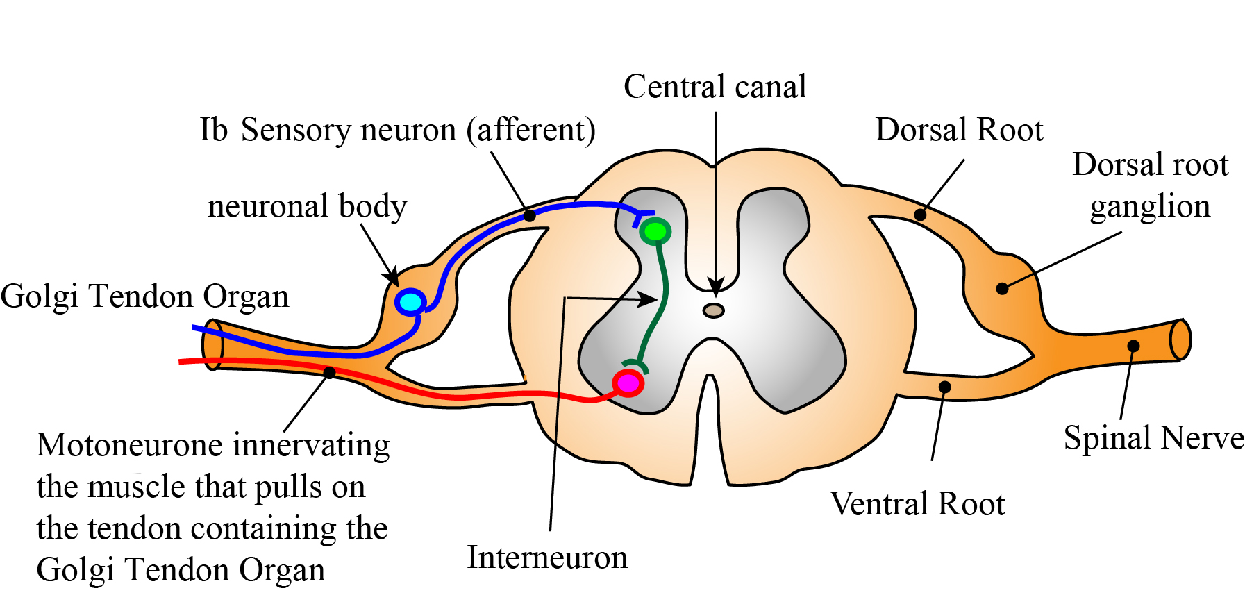
This reflex has some alternative names: the 'inverse myotatic reflex', or 'autogenic inhibition'. |
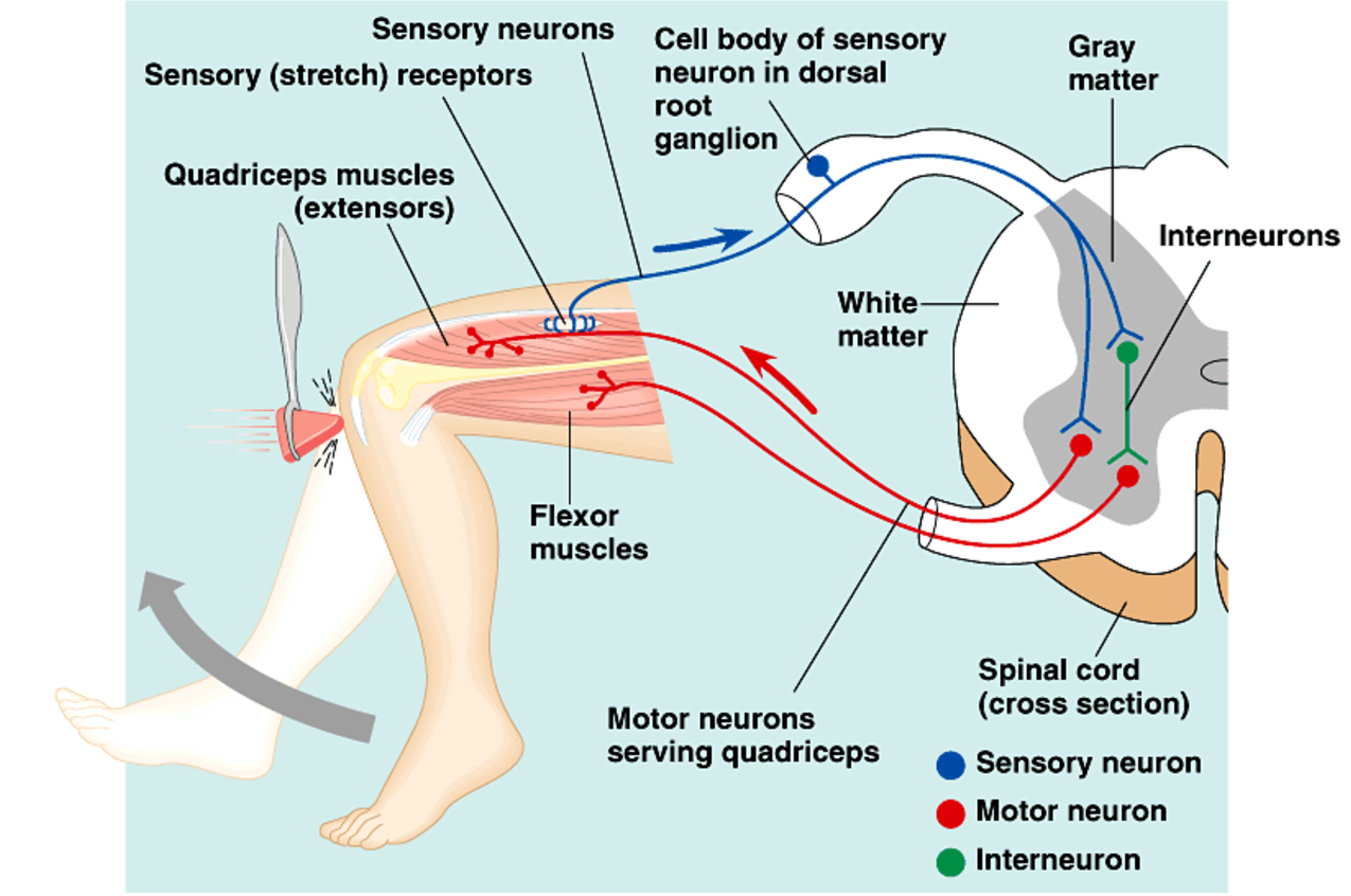
Disynaptic Reflexes: Reciprocal Inhibition In the knee jerk, contraction of quadriceps is accompanied by relaxation of the opposing flexor muscles. The contraction of quadriceps is mediated by a monosynaptic reflex (as stated above), and the relaxation of the antagonists is due to a disynaptic reflex. The relaxation of antagonists is mediated by an inhibitory transmitter released by interneurones - these are called the Ia inhibitory interneurones. Renshaw Cells : Recurrent Inhibition of Motoneurones The Renshaw Cell is an example of an interneurone that has an input from axon collaterals of alpha motoneurones before the axon enters the ventral horn. This excitatory input to Renshaw cells is medited by acetylcholine. Excitation of the Renshaw Cells inhibits the group of motoneurones gave rise to the recurrent axon collterals. More details in the parent webpage. |
 |
|
|
|
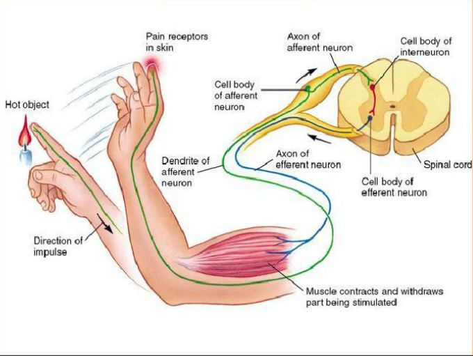
Polysynaptic Reflexes: the Flexion Withdrawal Reflex The flexor withdrawal reflex has a complex polysynaptic pathway that causes withdrawal of a limb from a noxious stimulus and maintain the withdrawn position. When injurious stimuli are applied to the skin, there is reflex withdrawal of the skin from the source of the injury. The sensory receptors in skin that mediate this reflex are known as nociceptors, a type of receptor that senses different types of noxious stimuli. Although pain is a normal accompaniment of this process, the pathway involved in this reflex response is entirely within the spinal cord, and the withdrawal responses occurs without the sensation of pain because the spinal cord is not directly involved in perception. The reflex responses is called the flexor withdrawal reflexand involves:
|
 |
|
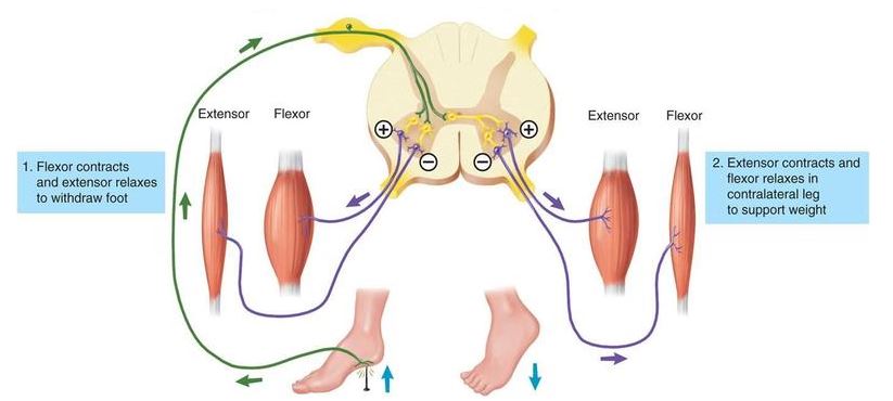
Polysynaptic Reflexes: the Crossed Extensor Reflex This reflex occurs as part of the flexor withdrawal reflex and ensures that extensors in the opposite limb contract so as to maintain the upright posture and support the weight of the whole body alone. The pathways that accomplish this are polysynaptic and cross the midline of the spinal cord. When nociceptors in the opposite limb elicit a flexor reflex, the crossed polysynaptic pathway excites extensor motoneurones on the opposite side of the cord. As in the flexor reflex, reverberating circuits are involved in the prolonged excitation of extensor motoneurones, so as to maintain support for the body while the opposite limb is flexed. |
 |