Close

Autonomic Nervous System : the Gastro-Intestinal Tract


The Enteric Nervous System

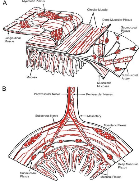
Within the gut wall there is a network of neuronal connections which senses chemical and physical changes in the gut lumen and in the gut wall, and causes reflex changes in the motility and secretion of the tissues.  These local (intrinsic) reflexes include peristalsis. These enteric neurones communicate widely with each other and regulate digestive system activity.The intrinsic plexuses are located between the mucosa and the circular muscle (the submucosal plexus), and between the circular muscle and the longitudinal muscle (the myenteric plexus).

The postganglionic neurones of the parasympathetic nervous system occur within the eneteric plexuses and behave as though they are the effector neurones for intrinsic reflexes, as well as the final effector neurones in the parasympathetic pathway. The parasymapthetic pathway tends to facilitate the activity of gut.

Postganglionic neurones of the Sympathetic System terminate within the intramural ganglia, and generally inhibit the activity of intrinsic reflexes.

Myenteric or Auerbach's plexus  neurons innervate the muscle and secretory cells and contain different transmitters [acetylcholine, peptides like vasoactive intestinal peptide (VIP), substance P, enkephalin and nitric oxide or somatostatin]. These neurons may be excitatory or inhibitory - thiose containing nitric oxide synthase or somatostatin are generally inhibitory.

Submucosal (or Meissner's plexus) contains the cell bodies of sensory fibers connected with sensory receptors deep in the mucosa and tranmits sensory information about mucosal conditions to the myenteric plexus.

The arrangement of sensory and motor neurons within the gut wall allows for local or intrinsic nerve reflexes such as Peristalsis to occur.


Peristalsis and Segmentation Movements   Top

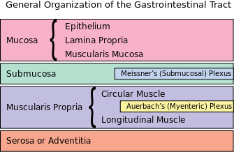
Peristalsis: a local reflex

Peristalsis is a propagated wave of circular muscle contraction that moves down the intestine and propels intestinal contents on to the next segment of bowel. The smooth muscle ahead of the propagated contraction also relaxes.

These events are mediated by intrinsic reflexes involving neurones within the gut wall plexuses. Sensory neurones respond to distension of the bowel, and these synapse on the neurones of the intrinsic plexuses, including effector neurones that release acetylcholine on the smooth muscle, causing it to contract. This final synapse in the pathway is sensitive to atropine, and is mediated by muscarinic receptors.

Segmentation Movements: generated by the spontaneous ativity of smooth muscle.

Segmentation movements cause intestinal contents to be mixed, without being propelled down the gut.  Segmentation movements are due to the spntaneous activity of smooth muscle; pacemaker cells and the interstitial cells of Cajal (ICC) may be involved in the genesis of segmentation movements.

These movements can be speeded up by vagal stimulation, and suppressed by sympathetic stimulation.


The effector neurones of the intrinsic plexus behave as though they were the final post-ganglionic neurones of the parasympathetic supply to the GI tract.

Chemoreceptors  that sense the osmolality, pH, and presence of substrates and products of digestion are also present, particularly in the upper duodenum, and help to regulate gastric emptying and the secretions of the pancreas and gall bladder.

The preganglionic fibres of the parasympathetic nervous system originate in the brain and in the sacral cord.  These fibres release acetylcholine and excite the postganglionic neurones, a process mediated by nicotinic recpetors.

The Vagus innervates the foregut, from the lower esophagus down to the transverse colon, whereas the parasympathetic supply to the lower gut is innervated from the pelvic nerves that originate in the sacral cord.

The postganglionic neurones of the sympathetic nervous system terminate on neurones of the intrinsic plexuses, and inhibit the reflex activities mediated by the plexus.

Postganglionic sympathetic neurones also end on the blood vessels, as elsewhere in the body.


Top Close


Actions of Parasympathetic and Sympathetic Nerves

The parasympathetic supply to the gut is carried in the vagus and pelvic nerves, arising from the medulla and sacral cord respectively.

The Vagus innervates the foregut, from the pharynx to the middle of the transverse colon, and the remainder is innervated by the pelvic nerves. Both influence motility and secretion.

The Vagus causes contraction of the smooth muscle of the gut (except for the smooth muscle sphincters), and causes secretion of acid in the stomach.

The Vagus also has some effects on secretion gastrin and of neuroendocrine tissues such as the islets of Langerhans.

The sympatheic supply to the gut is carried by the splanchnic nerves and the lumbare splanchnic nerves. The preganglionic fibres do not generally synapse in the sympathetic chain but traverse it to reach the caeliac and inferior mesenteric ganglia.

Postganglionic neurones arising in these ganglia supply the length of the intra-abdominal portion of the gastrointestinal tract. The foregut is innervated from the coeliac ganglion, and the hindgut from the inferior mesenteric ganglion. These fibres act on the intramural ganglia to inhibit their activity. They also cause the smooth muscle sphincters to contract, opposing the action of the parasympathetic nerves.


Actions of the Autonomic Nervous System on the Stomach   Top

The vagal efferent fibres that release acetylcholine cause increased gastric motility, increased secretion of acid and increase secretion of the hormone gastrin. These processes together encompass most of the processes involved in gastric digestion: the mixing of food with acid, and the slow delivery of the resulting chyme into the duodenum.

Some vagal fibres release NANC transmitters and are reponsible for inhibition of the gastro-oesophageal sphincter and as receptive relaxation in the stomach, when food enters the GI Tract.

The effects of the sympathetic are mainly vascular, playing a part in the regulation of blood flow to the stomach.

The colour of the gastric mucosa is known to change markedly when a person is under stress, and these changes are associated with sympathetic activity to the stomach.


Top Close


Parasympathetic and Sympathetic Reflexes

Autonomic nerve trunks contain visceral afferent nerve fibres as well as efferent axons, and these visceral afferents have importance in the mediation of reflexes and visceral pain. The majority of the neve fibres in the abdominal vagal tunk are in fact afferent, and these include mechanoreceptors and chemoreceptors that signal information about the state of the gut to the brain, and participate in reflex activity. In contrast to the local intrinsic reflexes mediated by the intramural plexuses (known as 'short' reflexes), the reflexes mediated by vagal afferents are known as 'long' reflexes or extrinsic reflexes.

The sympathetic nerves also contain afferent fibres, which are involved in visceral pain; some may also be involved in reflex functions. Vagal afferents appear not to be much involved in viscerall pain.

'In-series Tension receptors

'In-series Tension receptors' behave as though they were in series with smooth muscle in the gut wall, because they respond to stretch of the gut wall (caused by distension) and also to contractions of the visceral smooth muscle.

Chemoreceptors

Chemoreceptors in the gastrointestinal mucosa respond to changes in pH of the luminal contents.

Long Reflexes are reflexes mediated by afferent and efferent neurones that connect the gut to the CNS, and a coordination centre within the CNS.

Gastric distension activates the in-series tension receptors in the stomach wall, and the information is transmitted to the nervous system, where it contributes to the sense of satiety.

Those going to the rest of the colon, rectum and anus arise from the sacral spinal segments and are carried in the pelvic nerves. 

Stimulation of parasympathetic nerves can produce either stimulation or inhibition of smooth muscle but only stimulation of secretory cells.

Muscarinic cholinergic receptors are responsible for the stimulatory effects.

NANC transmitters are reponsible for some responses, such as receptive relaxation in the stomach.


Top Close


Oesophagus

The Upper 2/3 of the oesophagus contains striated muscle. Smooth muscle is present in the lower 2/3 of the oesophagus. This striated muscle is innervated by alpha motoneurones that have their cell bodies in the vagal and other motor nuclei in the brainstem, and in the sacral cord. 

Skeletal muscle tone is generated by impulses arising in the motoneurone cell bodies in the vagal nulcei and sacral cord . As elsewhere in the body, alpha motoneurones release the neurotransmitter acetylcholine which acts on nicotinic receptors on the endplates of the skeletal muscle fibres.

There is no intrinsic plexus innervation of striated muscle. 

Relaxation of skeletal muscle occurs by reducing the frequency of action potentials in the alpha motoneurones.

Secondary Peristalsis

The upper oesophagus also shows movements that resemble peristalsis. However the muscle is striated muscle and coordinated movements of this part of the oesphagus that move contents towards the somach can be elicitied by distension of the lumen.

The pathway for ths reflex involves a long reflex: sensory receptors in the upper oesphagus sense the presence of distension and send impulses to the medulla, where motoneurones innervating the striated muscle are activated. The skeletal muscle contracts in a coordinated muscle and drives the oesphageal contents towards the stomach.


Top Close


Rectum and Anus

The sphincteric mechanism of the anal canal involves an internal smooth muscle sphincter, and at least two sets of striated muscles, including the levator ani, which acts a a sling pulling on the back of the rectum, and the external sphincter.

The levator ani and external sphincter are innervated by alpha motoneurones arising within the sacral cord. The pudendal nerve motoneurones that contract these muscles originate form a region of the ventral horm known as Onuf's nucleus.

Distension of the rectum causes a reflex contraction of the colon and rectum and a relaxation of the anal sphincter, and this is the mechanism of defaecation.

The smooth muscle of the rectum is innervated by parasympathetic fibres that run in the pelvic nerves and cause contractions; these contractions are mediated by effector neurones that are part of the intramural plexus and are also postganglionic neurones.

The sympathetic innervation arises in the upper lumbar cord and synapses in the inferior mesenteric ganglion. It relaxes the smooth muscle and constricts the blood vessels.


Top Close

© HumanPhysiology.Academy 2014-2015