Surface Anatomy of the Cerebral Hemispheres
 * *
|
Lobes of the Cerebral Hemispheres Top |
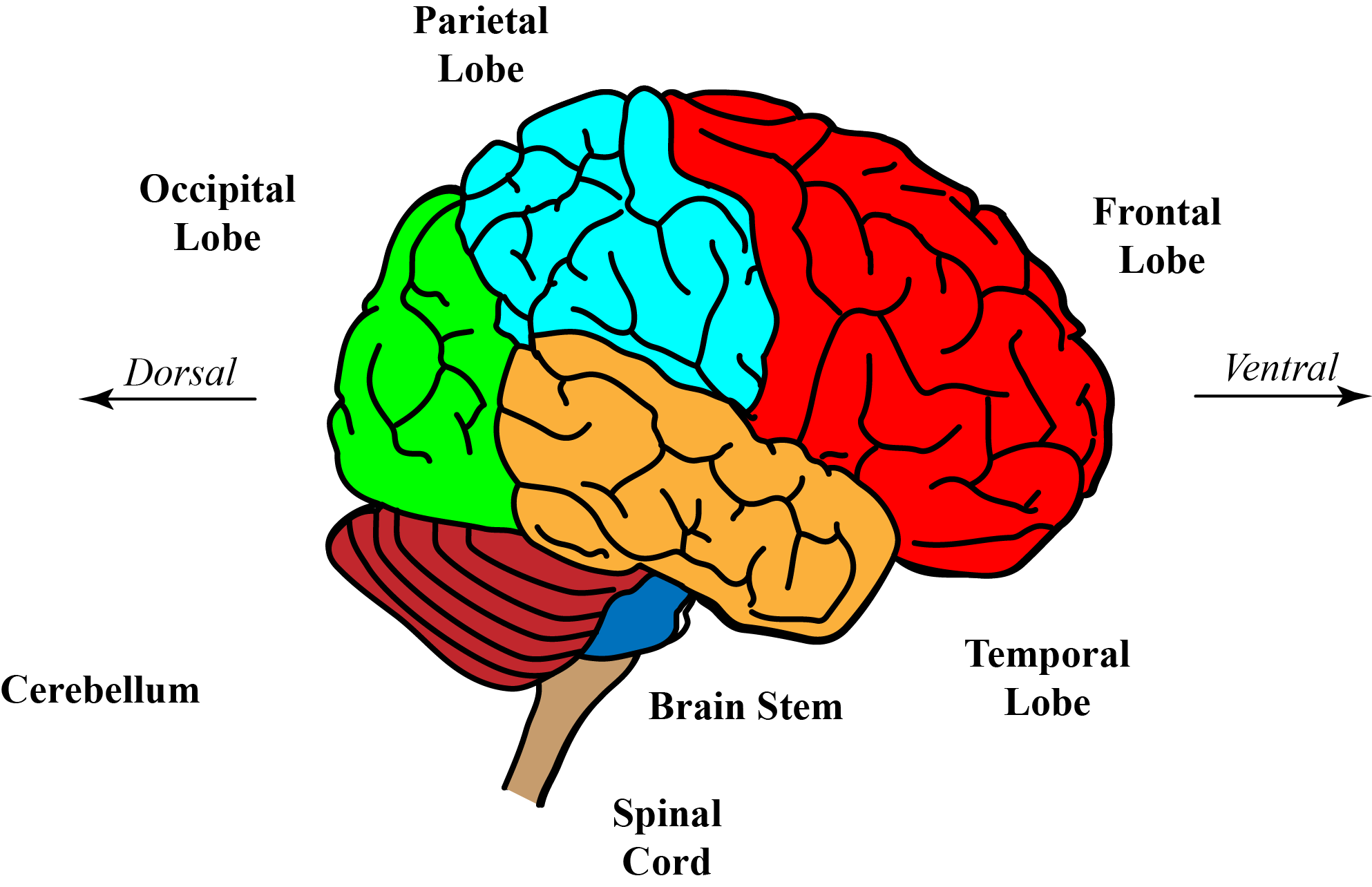
The surface of the cerebral hemispheres is convoluted and folded, creating some deep fissures which help to divide the cerebrum into lobes. These fissures include the central sulcus and the Sylvian fissure.
|
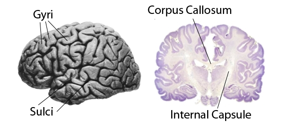
Some fissures are less deep than others, and are called sulci, and separate the surface folds which are called gyri. Generally speaking, the gyri all appear the same, i.e. grey folds on the surface of the brain. |
The central sulcus is the deep groove that separates the frontal and parietal lobes, and is a constant feature of the human brain. |
Prominent gyri are seen on each side of the central sulcus, and this fissure defines the pre- and post-central gyri on either side of the fissure. The precentral gyrus is concerned with fine control of movement, while the post-central gyrus is concerned with fine sensation. |
The cerebral hemispheres are divided into the frontal, occipital, temporal, and parietal lobes.
|
The central sulcus is the fissure that separates the frontal lobe from the parietal lobe. The Sylvian fissure separates the frontal lobes from the temporal lobes |
The frontal lobe is concerned with planning and the control of movement, while the parietal lobe is concerned with somatic sensation and integration of sensory stimuli. |
The occipital lobe is concerned with processing visual information, and the temporal lobe is concerned with hearing and speech
|
Internal Structure of the Cerebral Hemispheres Top
  * * |
The outer covering of the cerebral hemispheres is grey because it contains a high density of cell bodies, and is called the cerebral cortex. The cerebral cortex consists of six layers of cells. All parts of the cerebral cortex are basically similar in structure, but different areas of the cortex, known as Brodmann's areas, have some differences in the detail of arrangement of the cells. More information on the Cellular Structure of the Cerebral Cortex.
|
Below the cortex is white matter- axons that communicate with many other parts of the CNS, and other parts of the cerebral cortex. One important structure by which one hemisphere communicates with the other is a large band of axons called the corpus callosum.
The corpus callosum is the largest commissure in the CNS - the largest band of fibres crossing the midline. |
The coronal section of the brain (above) shows the corpus callosum and the internal capsule, which carries axons bertween the cortex and lower levels of the CNS. Sensory neurones carry infomation from the thalamus to the cortex within the internal capsule. Also Corticospinal neurones run through the internal capsuel form the cortex and reach the spinal cord. |
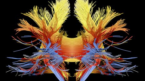
On the right you can see and artist's image of the white matter of the brain after the cortex has been removed, showing the axons that pass through the internal capsule and connect with the spinal cord in purple (at the bottom of the picture). |
The lobes of the cerebral cortex are highly interconnected by subcortical bands of white matter called association fibres.
|
Commissural fibers connect one hemisphere with the other, and the millions of commissural axons of the corpus callosum link the two hemispheres. |
Disconnection of the two hemispheres by surgical division of the corpus callosum results in a condition ('Split Brain') where the two sides of the brain may function independently, without coordination. |
In addition to the Frontal, Parietal, Occipital and Temporal lobes, there is a small lobe, the Insula, concealed in the depths of the lateral sulcus (Sylvian fissure -shown in red in the animation), between the frontal and temporal lobes. The Insula is concerned with autonomic functions and nociception. |
|
|
|
Other Features of the Cerebral Hemispheres Top
|
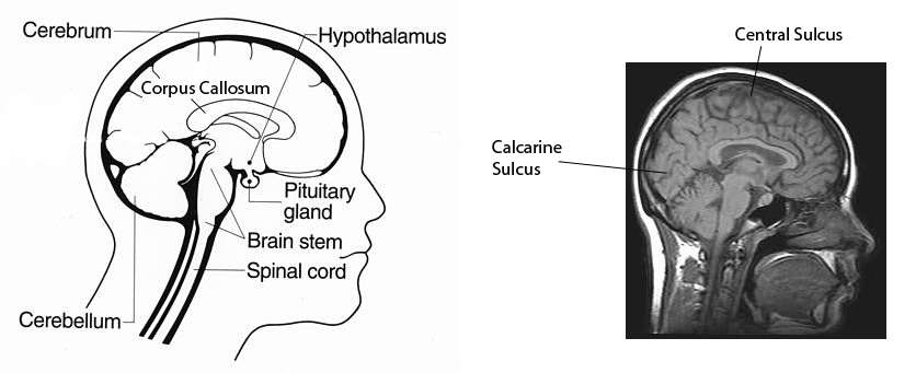
On the medial surface of each cerebral hemisphere the folding of the surface of the brain continues, and one major sulcus, the calcarine sulcus, is a constant feature on the medial surface of the occipital lobe. The calcarine gyri are part of the occipital lobe and are concerned with the analysis of visual signals. |
Another major feature of the medial surface of the brain is the Corpus Callosum, a substantial white band of nerve fibres that transfer information between the two hemispheres.
Many of these features can be seen by the use of Magnetic Resonance Imaging, which produces good images of the brain (right). |
Connections between Cortical Areas with Different Functions
There are also important connections in both directions between the cortex and the thalamus and basal ganglia. The thalamus is the main source of fibres entering the cortex from the deep nuclei of the brain.
Gross Anatomy of the Thalamus |
There are several main divisions of the cerebral cortex (see the diagram below):
- sensory cortex (somatosensory, visual, auditory)
- motor cortex (motor cortex, premotor cortex, frontal eye fields)
- association cortex (frontal, parietal, temporal) in between the other areas
|
The pre-central gyrus contains the cortico-spinal tract the pathway by which the motor cortex communictes with the muscles. This will be considered in more detail in the later |
The occipital lobe is concerned with vision and the analysis of visual images - shapes, colours, 3D interpretations etc.
More on the Visual pathway. |
The temporal lobe of the left hemisphere is concerned particularly with hearing and the interpretation of language, and communicates with areas of the frontal lobe concerned with speech via the arcuate fasciculus. |
Wernicke's area is concerned with interpretation of speech and language, whereas Broca's area in the frontal lobe is concerned with speech.
More on the auditory pathway, language and speech. |
|
The Cerebral Ventricles and the Blood Supply of the Brainstem and Cerebral Hemispheres Top
|
|
The brain starts life as the neural tube, built around the central canal. As the forebrain develops, large amounts of neural tissue develop round the central canal, which itself changes shape and has large expansions in its size in different ares of the brain.
These expansions of the rostral end of the central canal are known as ventricles.
Because the amount of neural tissue in the forebrain is so large it requires a large blood supply, and this is provided by the cerebral vessels.
The anatomy of the Cerebral Ventricles and Vessels is considered here |
|
|
| The Ventral Surface of the Brain Top |
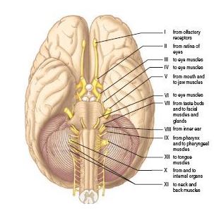 * *
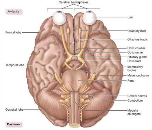
The diagrams show the two cerebral hemispheres, and the arrangement of the cranial nerves.
|  * *
|
An important marker on the ventral surface of the brain is the optic chiasma
at the anterior end of the hypothalamus, in front of the pituitary stalk. |
Half of the optic nerves axons from each eye cross to the other side of the brain in the optic chiasma, and pass
backwards, lateral to the mammilary bodies, in the optic tract. |
The optic tract axons relay within the lateral geniculate body (nucleus) which is part of the thalamus. Axons of the neurones of the lateral geniculate nucleus terminate mainly in the visual cortex. |
The neurones of the lateral geniculate nucleus send their axons to the peri-calcarine visual cortex for the analysis of visual signals..
Gross Anatomy of the Visual Pathway
|
The temporal lobe is separated from the frontal lobe by the Sylvian Fissure.
|
The connection between each cerebral hemisphere and the brainstem is the cerebral peduncles of the midbrain. |
The Olfactory Bulb is a relay nucleus for the sense of smell, and its axons project backwards to the orbital cortex- the ventral surface of the frontal lobe. |
The cerebellum is attached to the brainstem, and within the skull there is a fibrous membrane, the tentorium, which separates the upper surface of the cerebellum from the occipital lobe of the cortex.
|
|