Close

The Ventricular System and Blood Supply of the Brain


Ventricular System of the Brain

Cerebrospinal Fluid

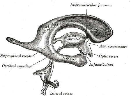
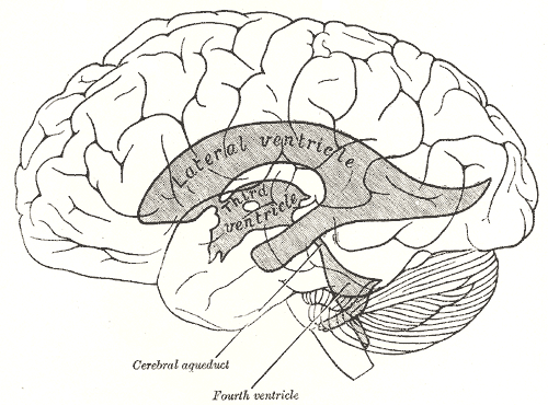
The brain and spinal cord float in the cerebro-spinal fluid (CSF), a salty fluid within the meninges. CSF is also present in the central canal of the spinal cord, in the space between the brainstem and cerebellum (fourth ventricle), in the lateral ventricles within each cerebral hemisphere and in channels that connect them (the aqueduct and third ventricle).

The nervous system develops from a tubular structure in the embryo called the neural tube. The central cavity of this tube remains in adults as the central canal of the spinal cord.

Cerebral Ventricles

In the brainstem the canal opens out into the cisterna magna and fourth ventricle.

In the midbrain it can be seen as a CSF-containing tube in the aqueduct, which expands into the third ventricle at the level of the thalamus.

The lateral ventricles are extensions of the third ventricle, connected by channels, the interventricular foramina, sometimes called the foramina of Monro, named after the Edinburgh anatomist.

*


*

*

Gray's Anatomy
Gray's Anatomy
www.obimages.net

Top Close


The Blood Vessels of the Brain

The brain has a high rate of metabolism and needs a high rate of blood flow to supply the oxygen for aerobic metabolism. The blood passes to the brain through the internal carotid and vertebral arteries on both sides.

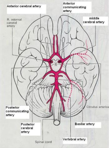
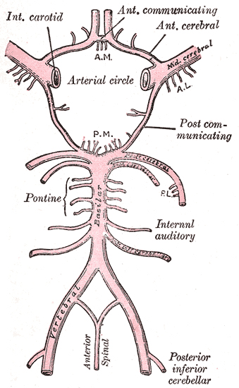
There is an important anastomosis between these vessels at the Circle of Willis and major cerebral arteries enter the substance of the brain from this structure.

The middle cerebral artery is particularly important in humans, as it is this artery that often becomes obstructed or haemorrhages, causing stroke. In this condition the neurones and axons that connect the cerebral cortex to the opposite side of the body are deprived of oxygen, resulting in paralysis and sensory loss.


The Arteries of the Cerebral Hemispheres and Brainstem   Top
Arteries

The Arterial Blood Supply to the brain comes through the right and left internal carotid arteries and the basilar artery (formed by the union of two vertebral arteries) that join together at the base of the brain to from the circle of Willis.

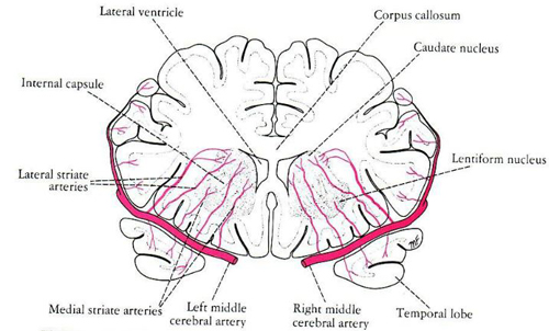
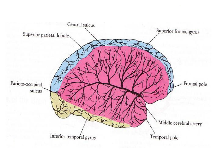
A number of arteries arise here to supply the cerebral hemispheres with blood; the middle cerebral artery is of particular importance in that it supplies blood to deep areas of the hemisphere and the areas of cerebral cortex concerned with sensation and motor control. 

The middle cerebral artery is of particular importance because it is commonly involved in the pathogenesis of stroke.


This coronal section shows that the middle cerebral artery originates from the Circle of Willis and sends branches through the striatum to the internal capsule and thalamus.

*

The distribution area of the middle cerebral artery supplies major areas of cerebral cortex, especially areas concerned with sensation, fine motor control and speech. Hence all of these are commonly affected if the artery becomes thrombosed or leaks blood into the surrounding tissues.

Tooltip trigger goes here.


*

Major Vessels supplying the brain

*

The Circle of Willis

Wikipedia
umqaa.blogspot.com
Docstoc

Top Close

The Venous Drainage of the Brain   Top

Veins.  

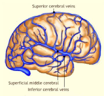
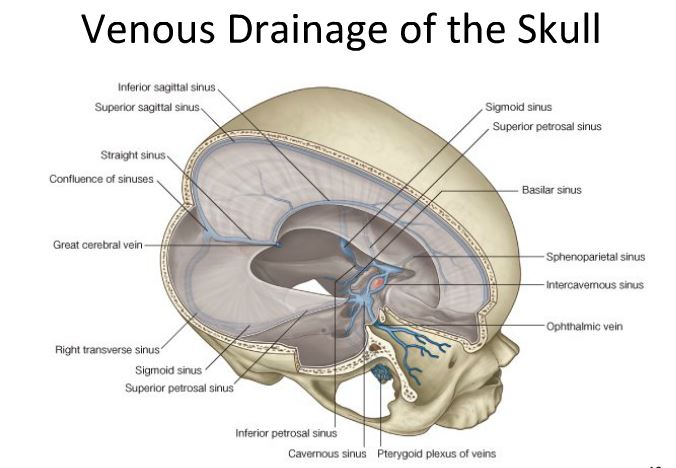
Venous blood from the brain enters the cerebral veins which converge on a series of sinuses that feed into the internal jugular vein.

The main veins on the surface of the brain are shown opposite.

The main venous sinus is the superior sagittal sinus which carries blood in the midline of the skull backwards towards the transvers and sigmoid sinuses which feed into the internal jugular vein.


Cavernous Sinus Thrombosis

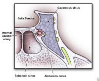
Veins from the eye and face drain into the cavernous sinus and then into the internal jugular vein. It should be noted that the cavernous sinus, because of its anatomical relationships to major nerves and other structures, can cause major disability if it becomes thrombosed.

In patients with cavernous sinus thrombosis, headaches often start within five to 10 days of developing an infection in the face or skull. Normally this infection would be treated with antibiotics, but if not other sympatoms develop. These include swelling, bulging and redness of the eyes, eye pain, double or blurred vision, and difficulty moving the eyes and the eyelids. Unless treated,patients with cavernous sinus thrombosis become increasingly drowsy and eventually fall into a coma.

*

*

The cavernous sinus is close to, but outside the pituitary fossa, and has relationships to the internal carotid artery and some major nerves innervating the eye and face.

The diagram shows the major veins within the skull, which drain into the internal jugular vein.

These larger veins taking blood from the brain and skull are protected the strong fibrous membranes such as the falx cerebri and the tentorium.

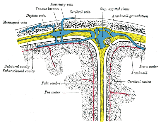
The superior sagittal sinus occurs at the top edge of a fibrous membrane that separates the two cerebral hemispheres, called the falx cerebri, which is continuous with the dura mater. Cerebral veins cross the subarachnoid space to enter the sinus.

*

*
emedicine.medscape.com
docstoc.com
Churchill Livingstone 2009 and pixshark.com
Gray's Anatomy

Close

© HumanPhysiology.Academy 2014-2015