 * * |
|
|
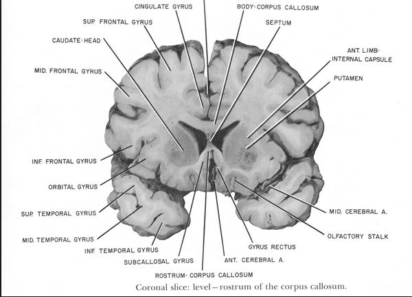
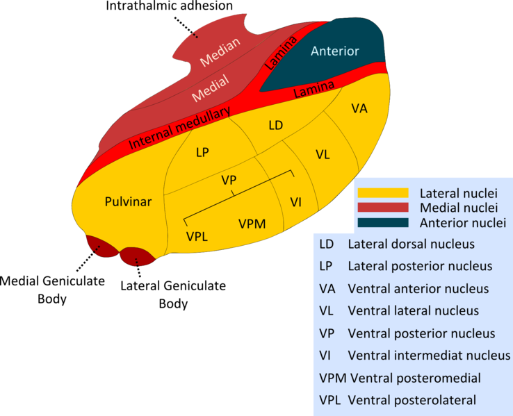
The thalamus consists of two large groups of nuclei, one on each side ot the third ventricle. The structure is approximately the size of a walnut and many of the axons of these nuclei project to the cerebral cortex. The two sides of the thalamus are separated by the third ventricle: in some individuals the sides of the third ventricle touch each other in a structure knon as the interthalamic adhesion; however no axons cross from one side to the other at this site. So each side of the thalamus is concerned strictly with sensory information from the contralateral side of the body, and objects in the contralateral visual and auditory fields. Much of the thalamus is concerned with processing sensory inputs and relaying their messages to the cortex. On the outer surface of the thalamus is a band of white matter called the internal capsule. This carries many of the axons arising within the thalmus to the cerebral cortex.; it also contains axons connecting the cerebral cortex with other parts of the CNS including the spinal cord. Theneurones of the Lateral Geniculate Nucleus (or Body) send their axons to the visual cortex in a fan-shaped arrangement of axons called the optic radiation; the axons arising from the lateral geniculate nucleus spread out to reach a large area of cortex around the calcarine fissure in the occipital lobe. Below the thalamus is the hypothalamus, a small area that regulates many physiological functions. | |
|
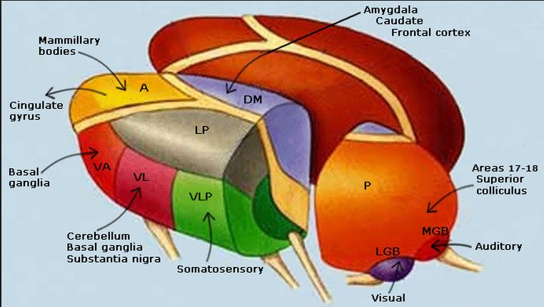
Different areas of the thalamus are separated by bands or sheets of myelinated nerve fibres called laminae (see below). At the posterior end of the thalamus is a large area called the pulvinar, and two important nuclei - the lateral geniculate body (or nucleus, LGN), and the medial geniculate body (or nucleus, MGN). The lateral geniculate nucleus is a relay station on the visual pathway, and the medial geniculate has a similar function for hearing. The ventro-basal complex of the thalamus consists of two main regions: the medial and lateral (VPM nucleus ventralis posteromedialis and VPL nucleus ventralis posterolateralis ) are concerned with somatic sensation of all types. The spinothalamic tract transmits information to the thalamus about pain, temperature, itch and crude touch. This area is the VPL area (nucleus ventralis posterolateralis ). There are two main inputs to VPL: the lateral spinothalamic tract, which transmits pain and temperature, and the anterior spinothalamic tract, which transmits crude touch and pressure information to these nuclei. The ventro-basal complex also handles the inputs from the dorsal column nuclei concerned with touch and proprioception. The anterior thalamus is concerned with emotion and memory, and recieves information from the mamillary bodies via the mammillo-thalamic tract, which consists of the mamillary body and fornix. This pathway connects with the cingulate gyrus and the hippocampus, concerned with emotion and memory. The thalamus also has a role to play in relaying signals concerned with motor function from the basal ganglia and the cerebellum to the cortex (VA and VL). |
|
 |
 * * |
One of the main functions of the thalamus is concerned with sensation. The pathways of senses including touch, pain, vision and hearing all have their own distinct nuclei in the thalamus. These nuclei relay afferent information to relevant areas of the cerebral cortex. The nuclei concerned with the individual sensations are: Somatic Sensations : Ventro-Lateral-Posterior Nuclei (VLP):
Vision: the Lateral Geniculate Nucleus:
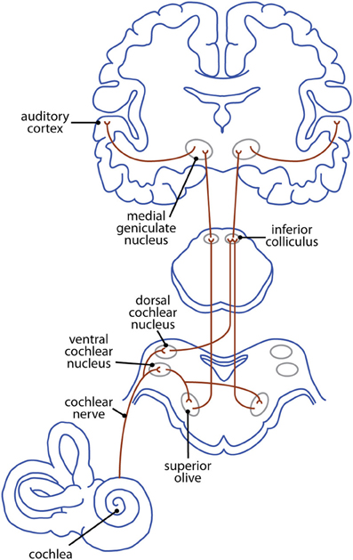
Hearing: the Medial Geniculate Nucleus
|
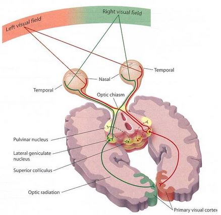
This diagram of the visual pathway shows the central role of the lateral geniculate nucleus in processing visual information. |
The Auditory Pathway The auditory pathway is complex as shown in the diagram opposite. The medial geniculate body is concerned with relaying the frequency and intensity of sounds to the auditory cortex; it also has multiple connections with the inferior colliculi. Information concerning the timing and intensity of information arising from both ears is also relayed to the cortex. There seem to be maps of sound frequencies within the inferior colliculi and the medial geniculate body (tonotopic maps, which correspond to the somatotopic map in the somatosensory cortex and the maps of the visual fields in the visual cortices). As for the visual pathway, the colliculi of the midbrain are concerned with eye movements. The superior colliculi are conerned with directing the gaze towards object in the visual field. The inferior colliculi are concerned with moving the eyes towards objects that are identified in auditory space. Primary Auditory Cortex The primary auditory cortex is essential for the perception of sounds, but central deafness is rare in humans because both cortices receive similar information, and it is rare for both to be injured by lesions. Eye Movements in relation to the direction of sound The inferior colliculi are concerned with directing the gaze towards objects that are identified in auditory space. Lower parts of the auditory pathway can still function however if the perception of sound is lost, so eye movements in response to sound can still occur in the absence of function auditory cortices. |
 * *
|
|
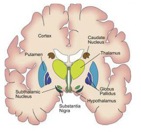
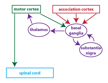
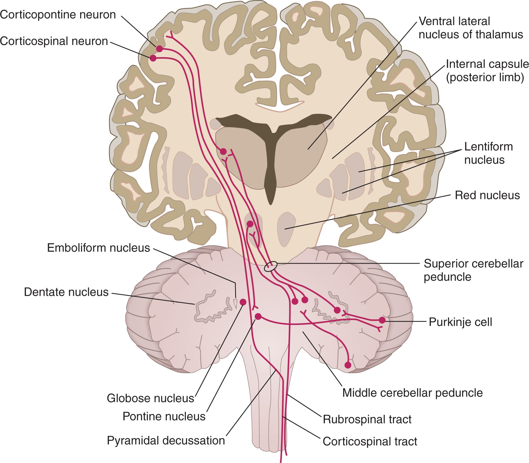
The thalamus relays signals concerned with motor function from the basal ganglia and the cerebellum to the cortex ; these functions are processed by the Ventro-Anterior and Ventro-Lateral Nuclei (VA and VL). The diagram opposite shows the thalamus and internal capsule,along with the cortex brainstem and cerebellum. The ventrolateral nucleus of the thalamus plays a crucial role in relaying to the cortex information arising from the cerebellum. In addition it shows the internal capsule and its important role in carrying axons of the corticospinal tract. The diagram opposite also shows the Lentiform nucleus (striatum) of the Basal Ganglia in cross section; this part of the basal ganglia also has important connections with the cortex and the thalamus. Below is a diagram that indicates the importance of the thalamus in connecting the output neurones of the basal ganglia ith the motor cortex.
|
|