Chapter 3 : The Forebrain and Somatosensory System

Brain: Contents Page

Functions of the Cerebral Cortex

Overview

The cerebral cortex has many functions including:

  • Primary Receiving Areas for sensations of vision, hearing and touch
  • Primary Motor Areas that execute movements
  • Language: Speech, and the interpretation of speech
  • Intelligence, Personality, Planning, Organization
  • Thinking, Perception, Interpretation of events

The lobes also have different functions:

  • Frontal Lobe - involved in planning, decision and problem solving.
  • Occipital Lobe - involved with vision.
  • Temporal Lobe - involved with memory, emotion, hearing, and language.
  • Parietal Lobe - receives and integrates sensory information.
  • The left and right hemispheres are not equally responsible for all functions (e.g.speech)
  • One hemisphere tends to be dominant e.g. right or left handedness.

Cortical Columns

All the main cortical sensory and motor areas contain columns of neurones with similar functions. However the properties of adjacent columns are slightly different, and sensations such as touch and vision use the slight differences between their receptive fields, and other properties, of adjacent cortical columns to enhance sensory discrimination.

Top

Methodology

Scientists have used different methods to find out which parts of the cerebral cortex do different jobs. Animal studies provided a strong basis for further wotk on humans. These include:

Brain damage: by studying animals or patients with brain damage, scientists have identified which parts of the brain are associated with different functions.

Electrical stimulation: Scientists have stimulated different parts of the brain with weak electrical currents and observed movements in the musculature or changes in behaviour. In humans, electrical stimuli to areas of the brain are known to induce movements, sensations, emotions, memories, etc. It is now routine to expose the brain under general anaesthesia, then let the patient wake up while maintaining a pain-free state using local anaesthesia to sensitive structures while the brain is exposed; in this situation, the patient can report the effects of electrical stimulation using electrodes that have been placed stereotactically into desired areas of the brain. If the motor area is stimulated, the patient makes an involuntary movement, and reports that the movement has occurred.. If the visual area is stimulated, they may see a flash of colour; if the motor cortex is stimulated there is a movement of some muscles.. Deep brain stimulation is sometimes used as a treatment of some neurological conditions.

MRI brain scans: Modern imaging methods such as MRI (Magnetic Resonance Imaging) scans provide images of brain structure and function in great detail. If humans perform a specific task during a scan, scientists can identify which parts of the brain are active when the task is carried out.

Recent advances in imaging techniques allow radiologists to monitor local blood flow (fMRI) and metabolism (PET scanning), both of which change when a part of the brain increases its activity. These are powerful techniques that have changed our understanding of the brain.

The results of all these studies indicate that different regions of the brain have different functions, some of which can be located in the following diagram:

Top


Sensory Areas of the Cerebral Cortex

The diagram shows the lateral surface of a cerebral hemisphere: different areas were given numbers by Brodmann on the basis of their cellular structure. There is a strong correlation between cellular structure and physiological function in the cerebral cortex.

Sensory areas

There is a general rule that one cerebral hemisphere handles sensory inputs from the opposite side of the body. This is true of the somatosensory cortex, and the visual system, where each visual area processes information relating to the opposite visual field of both eyes. The auditory cortex is an exception however, because localisation of sound requires comparison of the times of arrival of sounds at both ears; hence each auditory cortex receives inputs from both cochleas.

Sensory inputs to the cortex arise from the thalamic nuclei, including the medial and lateral geniculate bodies. There is a topographic map of one half of the body's surface on the contralateral somatosensory cortex, and a map of the contralateral visual fields of both eyes on the visual cortex. There is also a map of the cochlear basilar membranes on the auditory cortex, in keeping with the need to localise sounds.

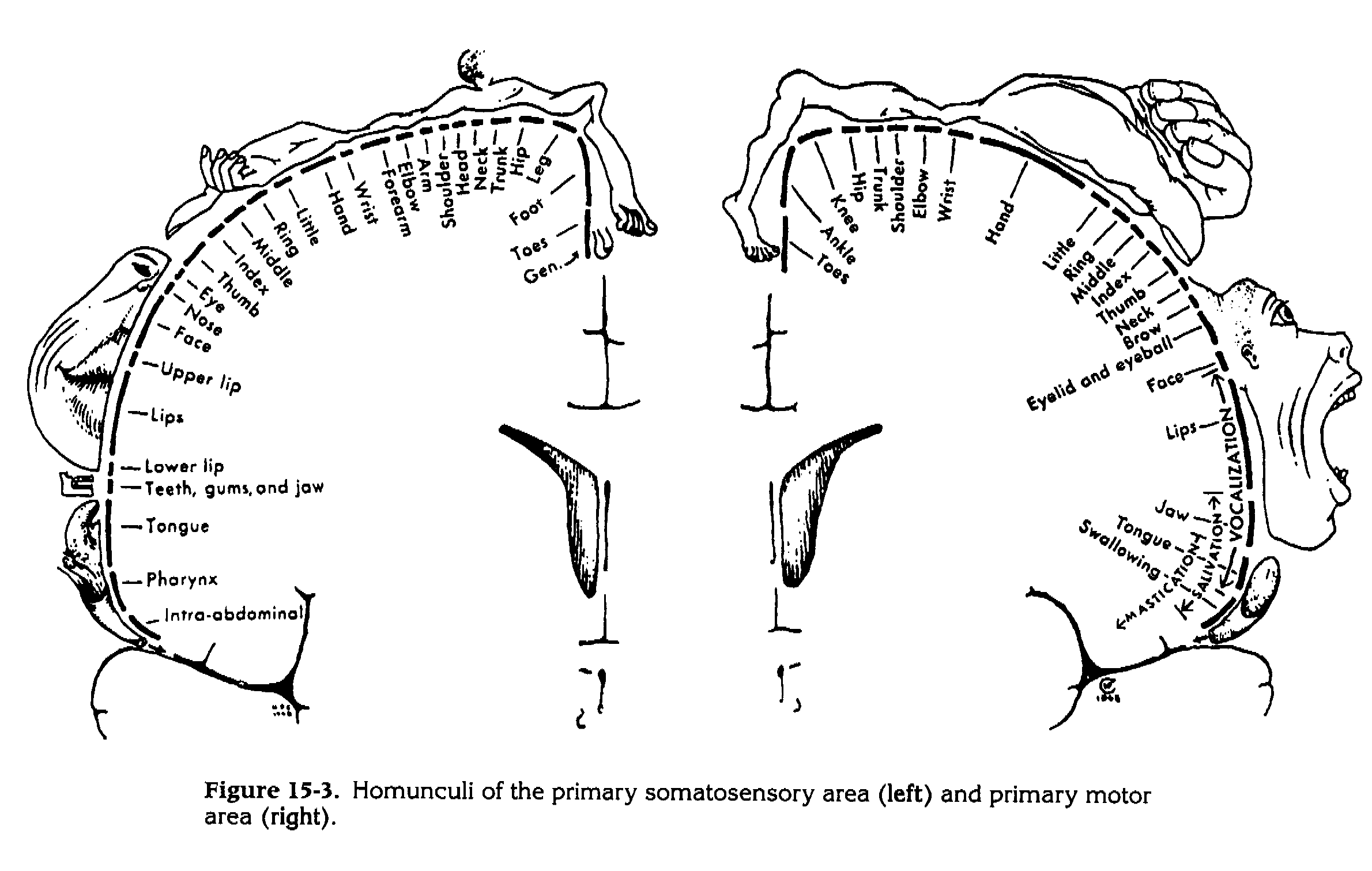
The cortical area given over to the fovea in the primary visual cortex is much larger that than given to the peripheral retinal fields; this is a result of the fovea having an extremely dense sensory innervation, allowing finer visual definition of objects focused on the fovea. The amount of cortex involved in processing signals from the fovea is correspondingly increased. Similarly, the sites on the body surface with greatest two-point discrimination (i.e. densest innervation) have the largest areas of somatosensory cortex. The map of the body surface on the cortex is therefore distorted, depending on the density of innervation of the skin, and this distorted map is called the homonculus.

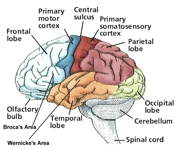
Somatosensory Cortex

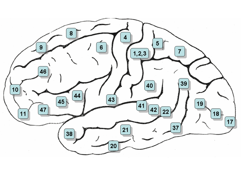
Primary somatosensory cortex (SI) is located in the post central gyrus (Brodmann's areas 1,2,3). This contains a somatotopic map, as does the thalamus, and in both structures the map of the body surface is distorted, depending on the density of sensory innervation in each part of the body. The hands, feet, genitalia and lips are the most sensitive areas (with the most dense innervation relative to the other areas) and these areas of the body surface have a relatively huge representaion on the cortex. As a result the sensory homunculus is grossly distorted with large areas of cortex given over to handling information arising in the most sensitive ares of the body. Damage to the sensory cortex results in decreased sensory thresholds, an inability to discriminate the properties of tactile stimuli or to identify objects by touch.

If a region of the body is amputated (such as a finger) there is reorganization of the primary somatosensory cortex with neurons that were previously activated by touching that finger now responding to stimulation of adjacent areas of skin..

In addition to SI, there is another representation of the body surface on the cerebral cortex - the secondary somatosensory cortex (SII) which is in Brodmann's area 40. It has inputs from both sides of the body - via SI and some less specific thalamic nuclei, but the map is less discrimintive. Some aspects of sensory experience are affected by damage to these areas.

*

The representation of the body surface on the primary somatosensory cortex, S1, (left) shows that areas with highest two-point discrimination have the greatest areas on the cortical map. Somatotopic Maps are found not only in S1, but also in the thalamus and dorsal column nuclei.

sca-germany_org

Top


Motor Areas of the Cerebral Cortex

Motor areas

The pre-central gyrus, the site of the primary motor cortex (MI, Brodmann's area 4), is involved in executing movements, and especially fine movements, e.g. of the fingers. The motor cortex in each hemisphere controls movements on the opposite side of the body. This is achieved by the corticospinal tract, which passes through the pyramids of the medulla, and is sometimes called the Pyramidal Tract. This area of cortex also sends axons to the nuclkei of the brainstem and to the thalamus and basal ganglia. These fibres send copies of the messages travelling down the Pyramidal tract to the cerebellum (via the brainstem) and all these structures work together to coordinate movements, posture and balance..

The somatotopic organization of the motor cortex has been defined using weak electrical stimuli that elicit defined movements. Movements of a specific joint tend to be represented (such as knee flexion) rather than movements of individual muscles. Damage to the motor cortex cause a weakness of the opposite side of the body, accompanied by spasticity. Clinicians call this an 'Upper Motoneurones Lesion'.

There are areas of the frontal lobe rostral to the motor cortex that are also involved in the control of movements, called the supplementary and pre-motor cortex. While the primary motor area executes movements because of the direct connection of the corticospinal tract with the alpha-motoneurones ('Lower Motoneurones'), the supplementary and pre-motor areas of cortex are involved in the selection of the necessary range of voluntary movements to achieve the objective.

The premotor cortex (Brodmann's area 6) is immediately anterior to the motor cortex much of its output is directed at the motor cortex; the premotor area also has lesser projections to the brain stem and the spinal cord. Area 6 lesions produce less severe weakness but greater spasticity than lesions of the precentral gyrus alone.

The basal ganglia also have an important role to play in planning and initiating movements.

Another area that projects to the primary motor cortex is the supplementary motor cortex (MII in Brodmann's area 6 on the medial side of the hemisphere), and appears to be involved in the intiation of movements as it, like the basal ganglia (with which it is connected), show electrical activity in advance of the primary motor cortex..

Eye Fields

Brodmann's area 8 is an area of the frontal lobe near the lateral end of the pre-central gyrus, and protruding into the frontal lobe. Electrical stimulation of this area causes coordinted horizontal movements of both eyes. The area has connections with structures involved in vision including the superior colliculi and the paramedian pontine reticular formation (PPRF) which are involved in vestibulo-ocular reflexes.


Top


Association Cortex

Association areas

Association areas organize and integrate sensory information of different modalities (touch, vision, hearing) into a coherent perceptual model of our environment. The parietal, temporal, and occipital lobes are all involved in associated these modalities and association fibres run beneath the cortex to integrate these inputs.

*

This diagram shows the patterns of short and long association fibres connecting different areas of the cortex. These are the means by which various functions are integrated to generate the higher level activity characteristic of the cortex

The association areas are adjacent to the primary receiving areas, e.g. the parietal lobe uses information from the somatosensory and visual cortices to guide the limbs, fingers and eye movements and generate complex movements requiring inputs from skin and eyes. Lesions of these areas leads to a failure to integrate sensory information. Areas behind the postcentral gyrus integrate visual and spatial inputs and are involved in perceiving an awareness of trajectories of moving objects. Unsurprisingly proprioception (awareness of the position of body parts in space) is also represented in this area..

The somatosensory association cortex (Brodmann's areas 5 and 7) receives inputs from SI and SII and lies directly behind the sensory cortex in the superior parietal lobes. Damage to this area causes tactile agnosia, an inability to recognize objects even though the objects can be felt. lesions of the parietal lobe also cause a neglect syndrome in which the patient is unaware of the opposite side of the body, and does not wash or clothe it.

pixshark.com

Top


The Executive Functions of the Frontal Lobes

Frontal Lobe Function.

The frontal lobes are extremely well developed in humans and these areas of cortex are involved in planning and executive functions. The prefrontal cortex is essential for attention, reasoning, judgment, planning and abstract thinking. Other areas on the inferior and medial surfaces of the lobe have links to the autonomic nervous system and limbic system; they are involved in mood, personality, reactions to events and control of impulsive acts.

*

In the 1860s railway supervisor, Phineas Gage,suffered an accident in which a metal rod penetrated his skull and frontal lobes. He survived and there is a classic description of the effects of this lesion:

"The equilibrium or balance, so to speak, between his intellectual faculties and animal propensities, seems to have been destroyed. He is fitful, irreverent, indulging at times in the grossest profanity (which was not previously his custom) manifesting but little deference for his fellows, impatient of restraint or advice when it conflicts with his desires, at times pertinaciously obstinate, yet capricious and vacillating, devising many plans of future operation, which no sooner arranged than they are abandoned... in this regard his mind was radically changed, so decidedly that his friends and acquaintances said that he was 'no longer Gage.'"

Based on these and other observations, neurosurgeons sometimes performed an operation called pre-frontal lobotomy. Surgical removal of parts of the frontal lobes as carried out in order to try and control certain behavioural disorders. These crude 'psycho-surgical' operations were soon banned, as it became clear that they were largely ineffective in achieving the desired result.

Speech

The role of the frontal and temporl cortices in the understanding of the spoken word and the formation of speech sounds are dealt with in the section on the Auditory System.

www.msmonographs.org

Top

Chapter 3 : The Forebrain and Somatosensory System

Brain: Contents Page

© HumanPhysiology.Academy 2014-2015