Vibration and Proprioceptive Senses: Receptors and Pathways |
|
Proprioception, sometimes called Kinaesthesia, is the awareness of the relative positions of parts of the body, particularly the limbs. There is also an awareness of the degree of muscular effort necessary for movements to be executed. Vibration Receptors in Muscle Proprioceptors in the limbs include muscle spindles, Golgi tendon organs and joint receptors. Muscle spindles are very sensitive to vibration and can induce changes in muscle tone when vibratory stimuli are applied to a muscle,- the tonic stretch reflex. |
Muscle spindles inform other neurons of the length of the muscle and the velocity of the stretch. This is particularly so for muscle that execute fine movements because muscle spindle density is highest in these muscle. There is also a high density of muscle spindles in the extensor muscle of the limbs that have to counter the effects of gravity and preserve posture. Muscle Spindles are also very sensitive to vibration and monitor the position of small joints Golgi Tendon Organs monitor the tensions at muscle-tendon junctions, and joint receptors sense the angle of each joint. |
|
Vibration Receptors in Skin Meissner's corpuscles also respond to vibration with an optimum frequence of around 30 Hz, whereas the highly sensitive Pacinian Corpuscles respond optimally to vibrations of around 250Hz. Sometimes the perception of vibratory stimuli is divided into two types on the basis of optimal frequency: frequencies of ~30Hz are referred to as 'flutter', and frequencies of ~200-300 are referred to as 'vibration'. Pacinian Corpuscles are found in deeper tissues such as the deeper layers of the dermis and connective tissues. Vibration can also be sensed by muscle spindles, whose afferents also contribute to the dorsal column-medial lemniscal system. All of these sensory receptors have myelinated axons. |
 * * The ways in which sensory receptors signal infomation is duscussed elsewhere |
|
Dorsal Columns and Dorsal Column Nuclei. Sensory modalities present in the dorsal column system are touch, pressure, vibration, and position sense. The sensory receptors involved are Pacinian Corpuscles, Meissner's Corpuscles and Mucsle Spindles. Position Sense (Kinaesthesia) involves afferent inputs form Muscles Spindles, Golgi Tendon Organs and Joint Receptors. The large myelinated axons involved in the senses of vibration and kinaesthesia have cell bodies in the dorsal root ganglia. The afferent axons enter the dorsal horn and their main collaterals pass up the dorsal columns of the same side of the cord to reach the dorsal root ganglia. |
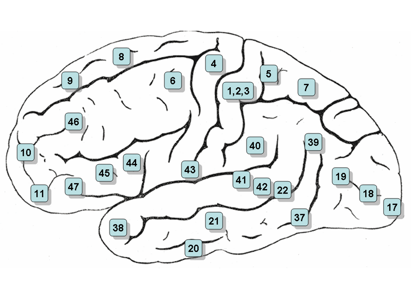
The neurones of the dorsal column nuclei have axons that project through the medial lemniscus to the ventro-basal nuclei of the opposite side of the thalamus. These axons cross the midline within the medulla, and form the medial lemniscus, which passes through the pons and the dorsal tegmentum of the midbrain to reach the thalamus. Somatosensory fibers are arranged in such a way as to preserve spatial information in their position, forming a map of the body surface known as a somatotopic map. Within this system, there are separate vibration and proprioceptive pathways (i.e.modality-specific private pathways) which reach the primary somatosensory cortex. |
