Close

The Anterolateral System : Alternative Nociceptive Pathways

The Anterolateral System.

The antero-lateral white matter of the cord contains not only the spino-thalamic tract, but other pathways arising from the dorsal horn and known to carry nociceptive signals.

Some of these axons project to the reticular formation and the midbrain, and together they form the Anterolateral System.

Some axons in the pain pathway project to the reticular formation (spino-reticular fibres) and to the periaqueductal grey matter of the midbrain.

Alternative Pain Pathways in the Anterolateral System

Nociceptive signals are distributed not only to the thalamus (via the spino-thalamic tract), but also to areas of the reticular formation of the brainstem, and to the periaqueductal grey matter of the midbrain.

All of these pathways originate within the dorsal horn and cross the midline near the central canal.

Spino-Reticular Tracts

The reticular formation of the brainstem is a hotchpotch of neurones that tend not to be grouped into well defined nuclei. They re the substance of the brainstem which surrounds the identifiable nuclei (such as the cranial nerve nuclei) and the fibre tracts that course through the pons and medulla. Some groups of neurones with indistinct boundaries are given names, such as the nucleus gigantocellularis reticularis and the midline raphe nuclei.

The left hand diagram shows the spino-reticular tract, whose axons terminate in the medulla and pons. Like the spino-thalamic tract these axons ascend the cord in the antero-lateral quadrant.

The axon terminals synapse in many parts of the the brainstem and the third order neurones of the pathways project to the thalamus.

*

Spino-Mesencephalic Tracts

On the right, the spino-mesencephalic tract is part of the antero-lateral system and ascends to synapse in the grey matter surrounding the aqueduct of the midbrain.

Referred Pain : Visceral Pain

Visceral pain is one of the strongest painful sensations known to man. It arises from internal organs as a result of occlusion of arteries (as in a heart attack), friction between layers of pleura or peritoneum (as in pleurisy or rebound tenderness in the abdomen), stretch of mesenteries, overdistension of viscera, or inflammation.

The spinal afferents that accompany the autonomic nerves are responsible for initiating visceral pain, but the second order neurones that project rostrally also carry afferent traffic from the skin. This is because of viscero-somatic convergence - both pathways (visceral and somatic) converge on the same neurones in the dorsal horn. A consequence of this is that activity in visceral afferents can be confused with somatic inputs within the same segments, and percieved as sensations within those somatic areas

Some examples:

  • afferents from the heart can induce sensations referred to the chest and arm (and sometimes in the neck)
  • afferents from the gall bladder can induce sensations in the abdominal skin, but also in the shoulder (because inflammation in that region can activate afferents of the phrenic nerve, whose origin is C4-5, which also innervates the shoulder)
  • afferents from the kidney and ureter can give rise to pain radiating from the small of the back to the groin.

*

Diagram of the convergence of somatic and visceral afferents on the neurones of the anterolteral system.


*

Tractotomy (Cordotomy). Neurosurgeons sometimes section the anterolateral system in an attempt to relieve chronic intractable pain by dividing the axons in the spinal cord.

This procedure can be successful, but breakthrough pain sensation can occur after a year or so, and one reason for this may be that there are alternative ascending pain pathways in the anterolateral system and other smaller pathways (such as the post-synaptic dorsal column pathway).

higheredbcs.wiley
medicguide

Top Close


Structure and Function of the Dorsal Horn in Relation to Nociception    Top

Rexed's Laminae

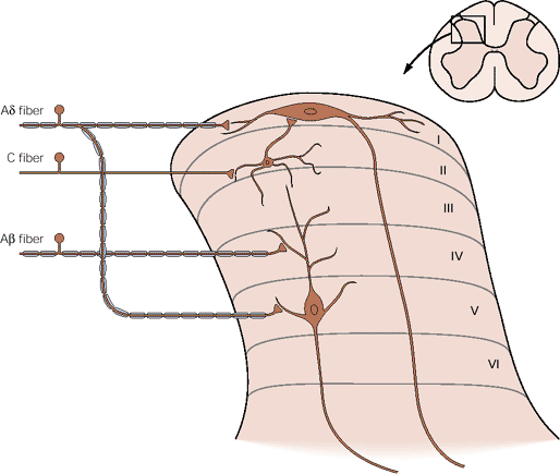
A neuroanatomist called Rexed looked at the structure of the grey matter of the spinal cord and divided it into columns of cells that were similar to each other in different segments.

His observations were based on the shape and size of the neurones, and he divided the grey matter into 10 (Roman X) layers and 6 of these are in the dorsal horn, numbered I to VI .

The classification has been useful in that the different lamina have neurones with different functions.

Lamina I was the marginal layer or zone.

Lamina II had a gelatinous appearance and is sometimes called the substantia gelatinosa.

Lamina IV and V had larger cells and this area has also been called the Nucleus Proprius

In Lamina VI another anatomist gave his name to Clarke's column, sometimes called the nucleus dorsalis.

In the ventral horn, lamina VIII and IX contain longitudinal columns of motoneurones

*

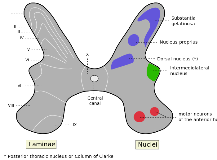
Lamina X is the area around the central canal.

In segments T1 to L2 and the first 4 sacral segments, the grey matter has an intermedio-lateral column of cells which give rise to autonomic neurones

The Dorsal Horn : Afferent Inputs

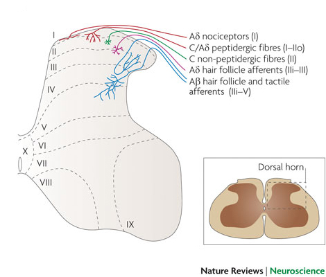
The dorsal horn is concerned with sensory functions, and different types of dorsal root axons originating from the skin and elsewhere synapse in different laminae of the dorsal horn. Some general principles apply: the superficial laminae are concerned with nociceptive sensations, and the deeper laminae process information from low threshold afferents. However some neurones in the depper laminae have inputs from nociceptive and tactile receptors.

Superficial Laminae

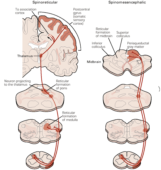
Laminae I and II are concerned with processing information from the nociceptive afferents which sense injury and inflammation in their receptive fields and are concerned with pain sensation. The neurones of lamina I, sometimes called marginal cells have long axons that project to the brainstem and thalamus.

Lamina II neurones (the substantia gelatinosa) is concerned with modulating the activity of Lamina I cells. Lamina II cells do not project outside the dorsal horn.

*


Nociceptive Transmission in the Dorsal Horn : The Gate Theory

*

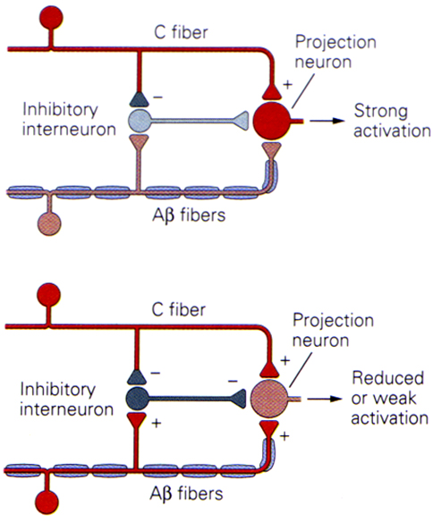
The schematic diagram above shows the concept of 'gating' in the dorsal horn of the spinal cord. It hypothesises that the balance of small and large fibre inputs to ascending neurones of the antero-lateral system modulates the activity transmitted by the ascending pathway.

The diagrams show a projection neurone, possibly equated with the marginal cells of Lamina I that project to the thalamus in the anterolateral system, and the arrangement of afferent neurones that could alter transmission in that pathway.

An inhibitory interneurone is shown (possibly equating to a Lamina II neurone) and the large and small diameter afferent inputs that regulate the activity of that cell can be seen.

The top diagram shows the response to stimulation of small fibres (nociceptors), whereas the bottom indcates the response to simultaneous activation of tactile and nociceptors.

The conclusion is that circuits of this type can modulate the activity of the antero-lateral system, depending on the balance of small and large diameter afferent inputs.

*

Deeper Laminae of the Dorsal Horn

Lamina IV and V receive inputs from afferents concerned with tactile sensation and with nociception. These cells are called 'wide dynamic range neurones' and project to the brain after crossing the midline and entering the lateral funiculi.


The Gate Theory suggests that the perception of pain depends on the balance of large and small fibre activity entering the dorsal horn, and that large myelinted fibre activity can reduce the onward transmission of nociceptive signals carried by unmyelinated axons.

There is now a lot of evidence that the pain signal can be modulated in the dorsal horn. Stimulation of large afferent fibres, by rubbing the skin, or by electrical stimulation of the skin (TENS) can reduce the perception of pain, and it is a common behaviour to rub an area of skin that has recently been hit by a hard object.

Dorsal column stimulation has been performed in humans using implanted electrodes; these stimulate a collateral of the large afferent neurones, and the impulse that passes antidromically down the cord appears to be able to reduce the perception of pain in ptients with chronic pain conditions. The suggestion is that this mechanism is identical to tht proposed in the Gate Theory.


Wikipedia

Top Close


Descending Control of Transmission in the Dorsal Horn   Top

Descending Control :

It is believed that the synapses in the dorsal horn can modulate the pain message. This can happen through to main mechanisms:

  • interactions between innocuous and nociceptive inputs to the dorsal horn (the 'Gate' Theory), and
  • descending pathays that terminate in the dorsal horn.

The diagram opposite shows some of the descending pathways that, when stimulated, inhibit nociceptive transmission in the dorsal horn.

They originate in:

  • the midline raphe nucleus of the medulla
  • the reticular formation of the brainstem
  • the periaqueductal grey matter of the midbrain
  • a region of the diencephalon known as A11
  • the Cingulate Gyrus of the cerebral cortex (part of the Limbic System)

The diagram also shows the importance of amines in influencing nociceptive processing in the dorsal horn.

Several transmitters are involved in the process:

  • 5-Hydroxytryptamine (Serotonin)
  • Noradrenaline
  • Dopamine

In addition there are interneurones in the dorsal horn that release enkephalins, which also have an inhibitory action at this site.


*


Top Close

© HumanPhysiology.Academy 2014-2015