The Main Pathway from the Motor Cortex to the Spinal Cord via the Corona Radiata, Internal Capsule and Brainstem |
|
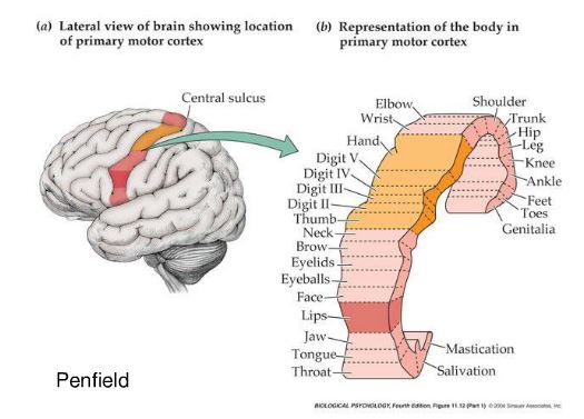
The motor cortex contains a map of the musculature of the body, and when we perform a voluntary movement, instructions are sent from this area of cortex to the motoneurones using a fast myelinated pathway - the corticospinal tract. The map of the musculature isn't a simple map of individual muscles, but involves groups of muscles concerned with the same movement. So movements rather than muscles are the nature of the signals emanating from the motor cortex.
|
|
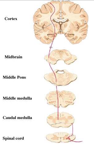
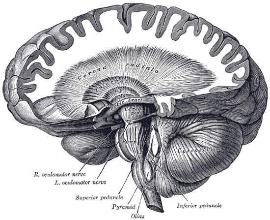
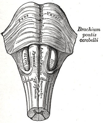
The Corticospinal Tract Top The main motor pathway, responsible for the execution of voluntary movements, begins in the motor cortex and is called the corticospinal tract. The size of this projection is large enough to be traced by dissection of the brain, and these nerve fibres pass through the corona radiata to the internal capsule, the cerebral peduncle, the pons and medulla. In the medulla 90% of the fibres cross to the opposite side in the decussation of the pyramids then pass down the lateral columns to reach the motoneurones on the side of the spinal cord opposite the motor cortex (the Lateral Corticospinal Tract). It is because of this decussation in the pyramids of the medulla that a lesion in the internal capsule causes paralysis on the opposite side of the body. Another name for the corticospinal tract is the Pyramidal Tract (because the axons pass through the pyramids). A smaller percentage of the descending fibres form the anterior corticospinal tract within the spinal cord, and pass down the cord ipsilaterally before crossing the midline at a segmental level to innervate the contralateral motoneurones. |
90% of corticospinal tract axons follow the path shown above. |
|
The Corona Radiata Top Nerve fibres connecting the cortex and lower structure in the CNS radiate out above the internal capsule forming a crown shaped structure when dissected. The Internal Capsule Top The internal capule is the main pathway the nerve fibres take between the cortex and the brainstem or spinal cord and is therefore of importance in the control of voluntary movements. The Internal Capsule is a collection of nerve fibres that pass in the narrow gap between the caudate nucleus and corpus striatum (putamen and globus pallidus) and then between the thalamus and corpus striatum to reach the cerebral peduncle. The Internal Capsule also carries the ascending fibres concerned with the sense of fine discriminative touch.
|
|
|
Corticospinal Tract in the Midbrain and Pons Top At the lower end of the internal capsule the corticospinal tract fibres enter the midbrain. They are in the superficial portion of the ventral surface of the cerebral peduncle. In the pons, the tract appears as bundles of fibres that pass through the substance of the pons and emerge in the ventral medulla near the midline. Medulla Top In the medulla a highly significant event occurs; the majority of the fibres cross over to the opposite side in an area called the pyramids. The name given to this crossing of the midline is the decussation of the pyramids. These axons continue into the spinal cord, mainly in the lateral funiculus to reach the motoneurones. A minority of the fibres continue into the spinal cord without crossing to the opposite side. These form the anterior corticospinal tract, and these fibres cross the midline to reach the motoneurones at a segmental level. |
 * * |
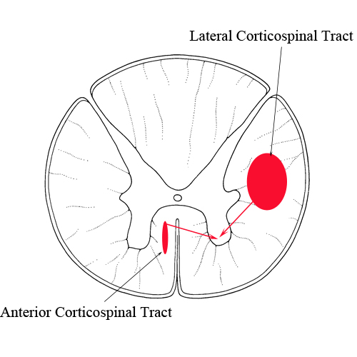
The Corticospinal Tract within the Spinal Cord Top In the spinal cord the majority of the corticospinal fibres (90%) travel down the cord in the lateral columns (having crossed the midline in the decussation of the pyramids) and make contact with motoneurones and interneurones in the ventral horn.
Anterior Corticospinal Tract A minor proportion (10%) of corticospinal tract axons do not cross the midline in the decussation of the pyramids, but continue to travel caudally in the anterior columns, before crossing over to the opposite ventral horn at a segmental level. These axons tend to make contact with motoneurones innervating the axial muscles.
|
|
