HumanPhysiology.academy

home  

Cerebro Spinal Fluid and the Blood-Brain-Barrier


A. Brain Metabolism

1. Brain metabolism

is very high; amounts to 15% of total body metabolism although it only weighs 2% of total body weight

2. Perfusion:

Perfusion is about 700-900 ml/min (or about 15% cardiac output)

3. Metabolism:

There is no anaerobic metabolism in the brain (no glycogen storage and no oxygen storage)

4. Aerobic metabolism:

There is only aerobic metabolism with O2 and glucose is immediately required from the blood. If the blood flow is stopped -> unconscious within 3-4 seconds.

5. Blood Flow Regulation:

Practically and totally autoregulated (pO2, PCO2 and pH dominated)

Blood flow shows active hyperaemia (very useful nowadays in brain imaging studies).

6. Blood Brain Barrier:

Barrier between blood and brain tissue. This barrier is caused by tight junctions between the endothelium in the brain capillaries (= anatomical BBB)

7. Physiological BBB:

  1. Easily transportable: lipid-soluble such as O2, CO2, water, anaesthetics.
  2. Medium transportable: electrolytes (charge!)
  3. Difficult transportable: proteins, non-lipid solubles

8. Biochemical BBB:

There are membrane-bound enzymes to destroy hormones and neuro-transmitters that may attempt to cross the barrier.


9.

The Blood brain Barrier is not constant throughout the brain. There are several areas where the barrier is less present or even absent leading to good communication between blood and nerve cells.


10.

One of these areas is the hypothalamus where nerve centers need to obtain information from the blood to regulate metabolic activities, water balance (ADH). Also, in (premature) babies, the BBB is not complete yet!


B. The Cerebro Spinal Fluid and the CSF System:


diagram of the cerebro spinal fluid system

1.

CSF is required to nourish brain tissue and to protect the brain from blows to the head (floating brain), together with the skull. However, because of the inertia of the fluid and the brain, there is a potential danger of the “contrecoup” (brain damage at the other side of the blow).

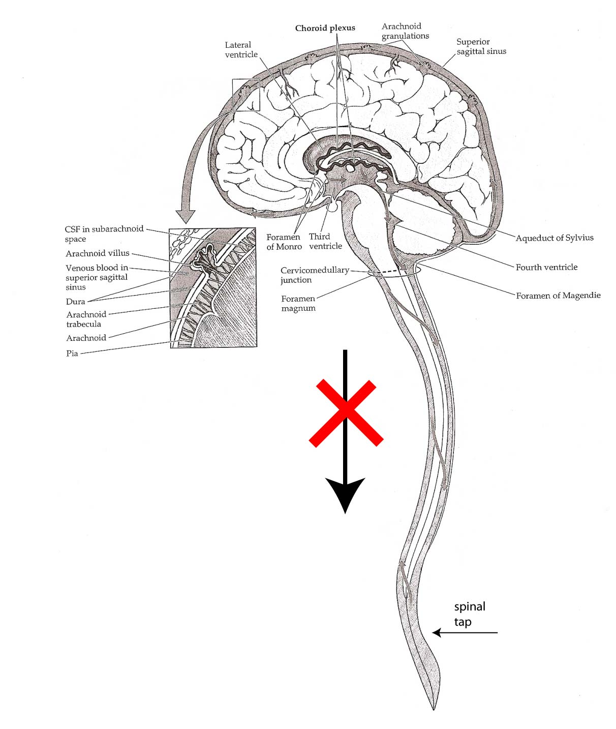
2.

CSF is produced by the choroid plexuses located in all four ventricles. Every day about 500 ml CSF is produced. The total quantity in a normal brain is about 150 ml; there is therefore a 3-4x daily renewal of all CSF.

3.

CSF flows from the Lateral ventricles, through the foramen of Monro into the Third ventricle and, through the Aqueduct of Sylvius, into the Fourth ventricle. This ventricle is connected though two foramina of Luschka and the foramen of Magendie into the Cisterna Magna. From there, fluid flows through the (sub)arachnoidal space and reaches the arachnoidal villi.

4.

The arachnoidal villi work actually asvalves between the brain fluid and the venous system. If the brain fluid pressure is higher than the local venous pressure (about 10 mm Hg), then CSF will flow into the veins and back to the heart.


5.

Measuring CSF pressure is done with a spinal tap. Buf before you do this, you MUST check for one thing!!

6.

The absence of papilledema. (indication of raised intracranial pressure -> risk of herniation of the brain stem -> acute death.


C. Hydrocephalus.

Literally:

too much water in the brain!

Non-communicating hydrocephalus:

Block in one of the canals so that CSF is still produced but cannot flow to the arachnoidal space. Often, the block is located in the aqueduct of Sylvius (atresia (closure) before birth or tumor at any age).

Communicating hydrocephalus:

fluid is NOT transported through the arachnoidal villi because these are blocked (caused by debris blockage from an infection, inflammation or bleeding). Therefore, the pressure is increased throughout the cranium

Therapy:

Shunt from the brain (ventricles) to another space in the body (peritoneal cavity, right atrium).


© HumanPhysiology.academy 2014